3U0O image
Deposition Date 2011-09-28
Release Date 2012-03-14
Last Version Date 2023-09-13
Entry Detail
PDB ID:
3U0O
Keywords:
Title:
The crystal structure of selenophosphate synthetase from E. coli
Biological Source:
Source Organism:
Host Organism:
Method Details:
Experimental Method:
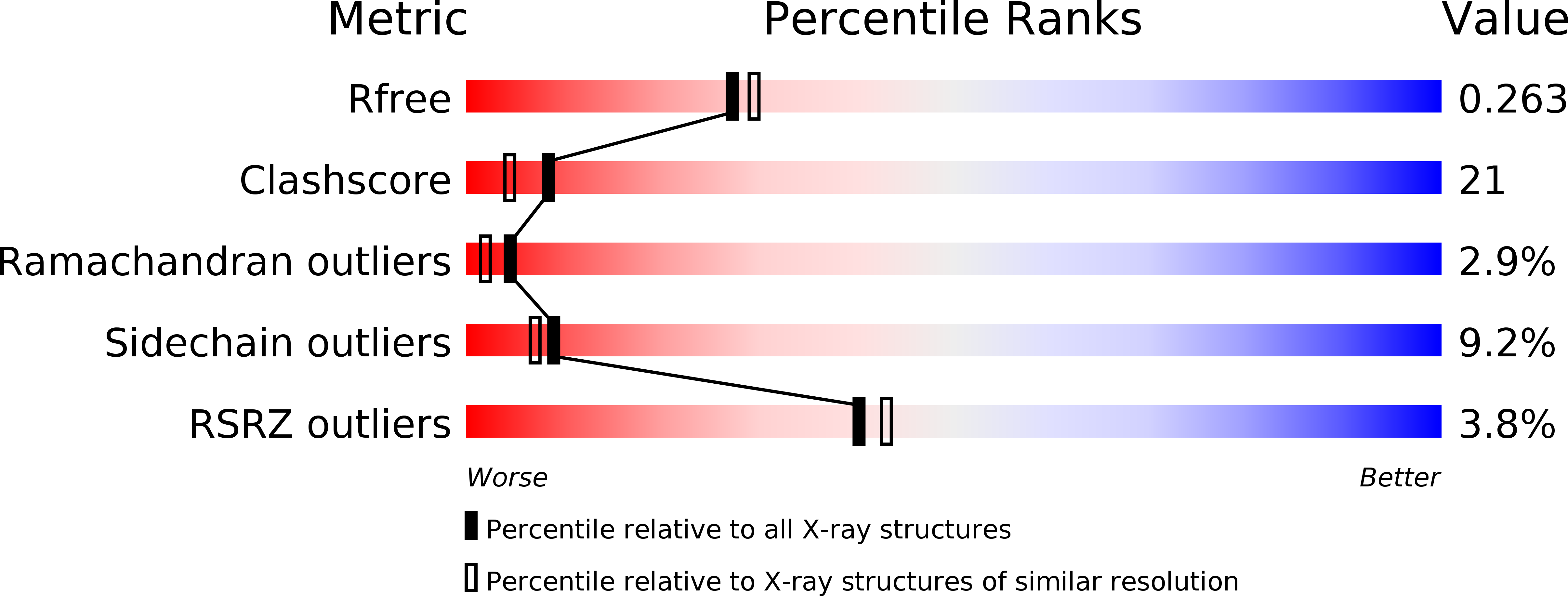
Resolution:
2.25 Å
R-Value Free:
0.26
R-Value Work:
0.22
R-Value Observed:
0.22
Space Group:
P 32 2 1
Macromolecular Entities
Structures with similar UniProt ID
Protein Blast
Polymer Type:polypeptide(L)
Molecule:Selenide, water dikinase
Gene (Uniprot):selD
Chain IDs:A, B
Chain Length:347
Number of Molecules:2
Biological Source:Escherichia coli
Ligand Molecules
Primary Citation
Structural insights into the catalytic mechanism of Escherichia coli selenophosphate synthetase.
J.Bacteriol. 194 499 508 (2012)
PMID: 22081394 DOI: 10.1128/JB.06012-11

Abstact

Selenophosphate synthetase (SPS) catalyzes the synthesis of selenophosphate, the selenium donor for the biosynthesis of selenocysteine and 2-selenouridine residues in seleno-tRNA. Selenocysteine, known as the 21st amino acid, is then incorporated into proteins during translation to form selenoproteins which serve a variety of cellular processes. SPS activity is dependent on both Mg(2+) and K(+) and uses ATP, selenide, and water to catalyze the formation of AMP, orthophosphate, and selenophosphate. In this reaction, the gamma phosphate of ATP is transferred to the selenide to form selenophosphate, while ADP is hydrolyzed to form orthophosphate and AMP. Most of what is known about the function of SPS has derived from studies investigating Escherichia coli SPS (EcSPS) as a model system. Here we report the crystal structure of the C17S mutant of SPS from E. coli (EcSPS(C17S)) in apo form (without ATP bound). EcSPS(C17S) crystallizes as a homodimer, which was further characterized by analytical ultracentrifugation experiments. The glycine-rich N-terminal region (residues 1 through 47) was found in the open conformation and was mostly ordered in both structures, with a magnesium cofactor bound at the active site of each monomer involving conserved aspartate residues. Mutating these conserved residues (D51, D68, D91, and D227) along with N87, also found at the active site, to alanine completely abolished AMP production in our activity assays, highlighting their essential role for catalysis in EcSPS. Based on the structural and biochemical analysis of EcSPS reported here and using information obtained from similar studies done with SPS orthologs from Aquifex aeolicus and humans, we propose a catalytic mechanism for EcSPS-mediated selenophosphate synthesis.

Legend

Protein

Chemical

Disease

Primary Citation of related structures