3U01 image
Deposition Date 2011-09-28
Release Date 2011-12-21
Last Version Date 2024-11-20
Entry Detail
PDB ID:
3U01
Title:
Crystal structure of onconase double mutant C30A/C75A at 1.12 A resolution
Biological Source:
Source Organism:
Rana pipiens (Taxon ID: 8404)
Host Organism:
Method Details:
Experimental Method:
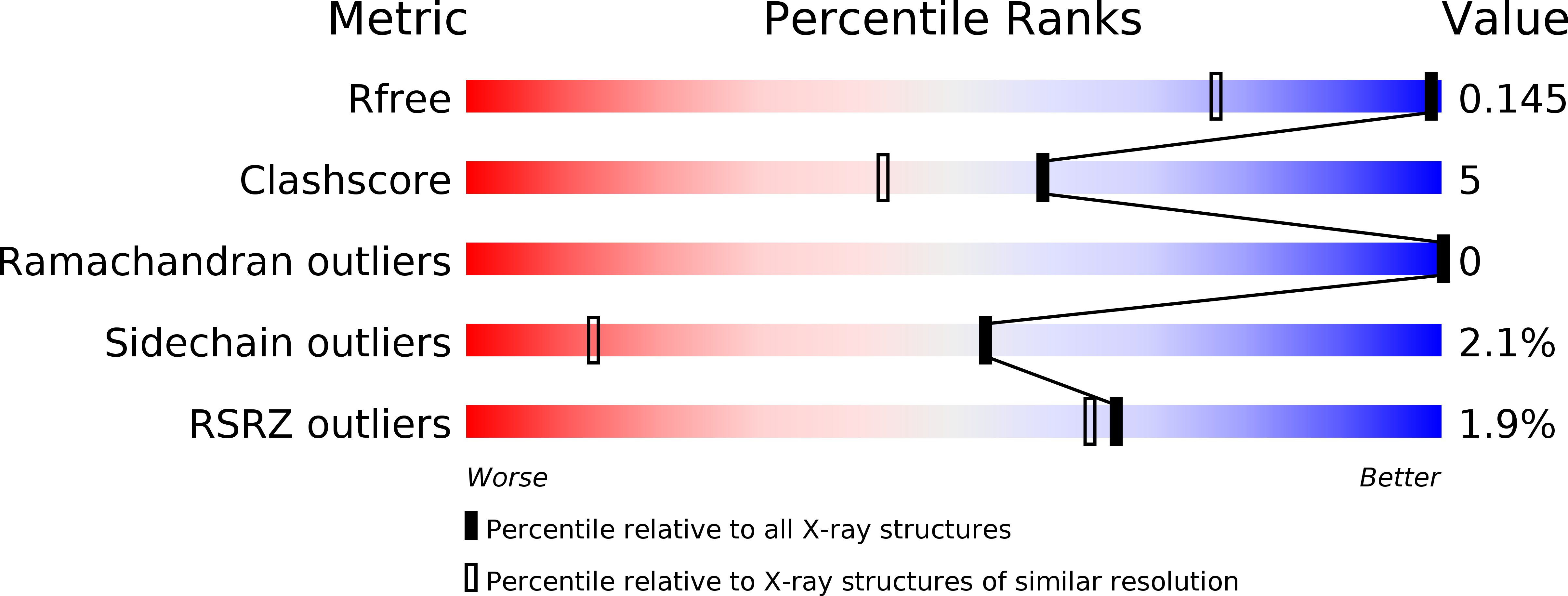
Resolution:
1.12 Å
R-Value Free:
0.14
R-Value Work:
0.13
R-Value Observed:
0.13
Space Group:
P 21 21 21
Macromolecular Entities
Structures with similar UniProt ID
Protein Blast
Polymer Type:polypeptide(L)
Molecule:Protein P-30
Mutagens:C30A/C75A
Chain IDs:A
Chain Length:104
Number of Molecules:1
Biological Source:Rana pipiens
Modified Residue
Compound ID Chain ID Parent Comp ID Details 2D Image
PCA A GLN PYROGLUTAMIC ACID
Primary Citation
Investigating the effects of double mutation C30A/C75A on onconase structure: Studies at atomic resolution.
Biopolymers 101 454 460 (2014)
PMID: 23996687 DOI: 10.1002/bip.22403

Abstact

The structure of onconase C30A/C75A double mutant has been determined at 1.12Å resolution. The structure has high structural homology to other onconase structures. The changes being results of mutation are relatively small, distributed asymmetrically around the two mutated positions, and they are observed not only in the mutation region but expanded to entire molecule. Different conformation of Lys31 side chain that influences the hydrogen bonding network around catalytic triad is probably responsible for lower catalytic efficiency of double mutant. The decrease in thermal stability observed for the onconase variant might be explained by a less dense packing as manifested by the increase of the molecular volume and the solvent accessible surface area.

Legend

Protein

Chemical

Disease

Primary Citation of related structures