3TYK image
Deposition Date 2011-09-26
Release Date 2011-10-12
Last Version Date 2022-04-13
Entry Detail
PDB ID:
3TYK
Title:
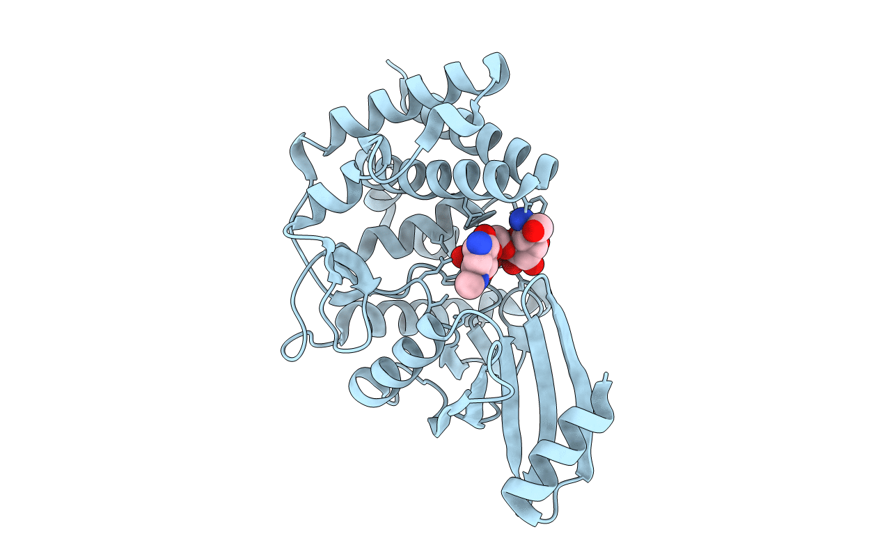
Crystal structure of aminoglycoside phosphotransferase APH(4)-Ia
Biological Source:
Source Organism:
Escherichia coli (Taxon ID: 562)
Host Organism:
Method Details:
Experimental Method:
Resolution:
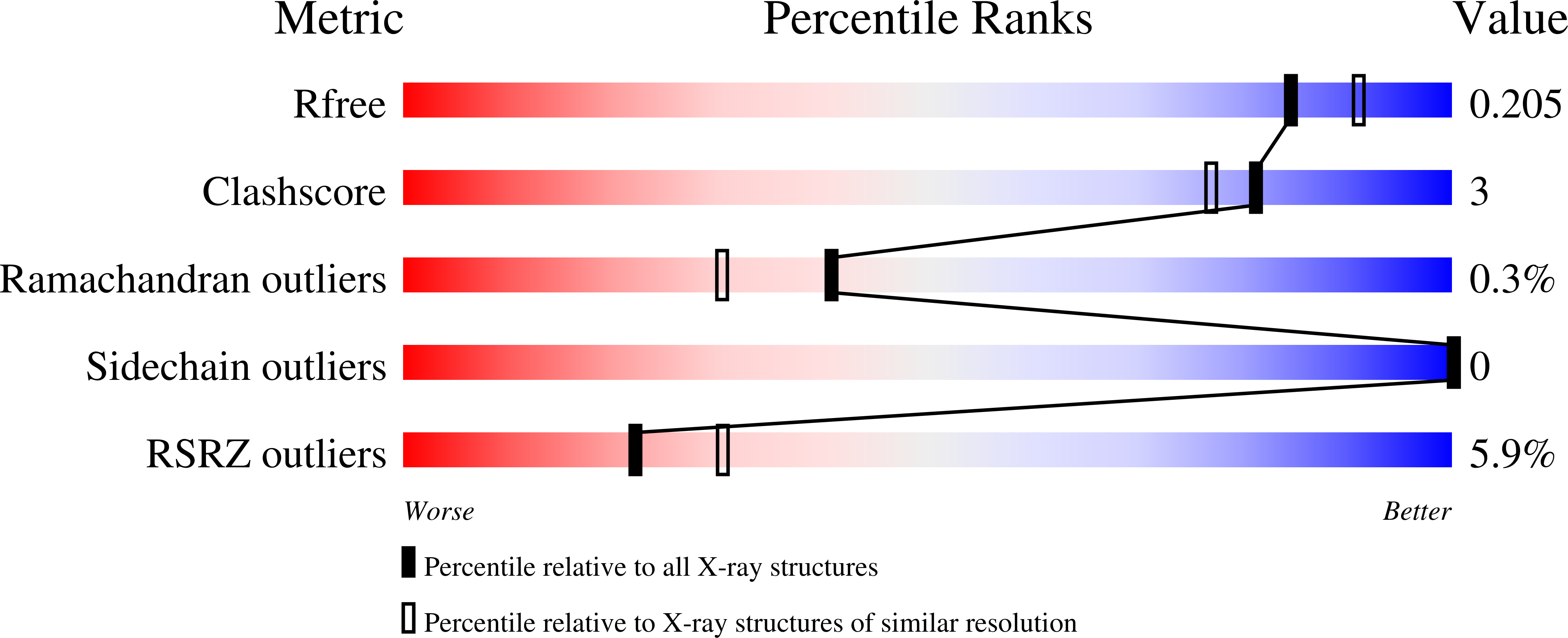
1.95 Å
R-Value Free:
0.20
R-Value Work:
0.15
R-Value Observed:
0.15
Space Group:
P 32 2 1
Macromolecular Entities
Polymer Type:polypeptide(L)
Molecule:Hygromycin-B 4-O-kinase
Gene (Uniprot):hph
Chain IDs:A
Chain Length:362
Number of Molecules:1
Biological Source:Escherichia coli
Modified Residue
Compound ID Chain ID Parent Comp ID Details 2D Image
CSX A CYS S-OXY CYSTEINE
MSE A MET SELENOMETHIONINE
Primary Citation
Structure and function of APH(4)-Ia, a hygromycin B resistance enzyme.
J.Biol.Chem. 286 1966 1975 (2011)
PMID: 21084294

Abstact

The aminoglycoside phosphotransferase (APH) APH(4)-Ia is one of two enzymes responsible for bacterial resistance to the atypical aminoglycoside antibiotic hygromycin B (hygB). The crystal structure of APH(4)-Ia enzyme was solved in complex with hygB at 1.95 Å resolution. The APH(4)-Ia structure adapts a general two-lobe architecture shared by other APH enzymes and eukaryotic kinases, with the active site located at the interdomain cavity. The enzyme forms an extended hydrogen bond network with hygB primarily through polar and acidic side chain groups. Individual alanine substitutions of seven residues involved in hygB binding did not have significant effect on APH(4)-Ia enzymatic activity, indicating that the binding affinity is spread across a distributed network. hygB appeared as the only substrate recognized by APH(4)-Ia among the panel of 14 aminoglycoside compounds. Analysis of the active site architecture and the interaction with the hygB molecule demonstrated several unique features supporting such restricted substrate specificity. Primarily the APH(4)-Ia substrate-binding site contains a cluster of hydrophobic residues that provides a complementary surface to the twisted structure of the substrate. Similar to APH(2″) enzymes, the APH(4)-Ia is able to utilize either ATP or GTP for phosphoryl transfer. The defined structural features of APH(4)-Ia interactions with hygB and the promiscuity in regard to ATP or GTP binding could be exploited for the design of novel aminoglycoside antibiotics or inhibitors of this enzyme.

Legend

Protein

Chemical

Disease

Primary Citation of related structures