3TXM image
Entry Detail
PDB ID:
3TXM
Title:
Crystal structure of Rpn6 from Drosophila melanogaster, Gd(3+) complex
Biological Source:
Source Organism:
PDB Version:
Deposition Date:
2011-09-23
Release Date:
2011-12-14
Method Details:
Experimental Method:
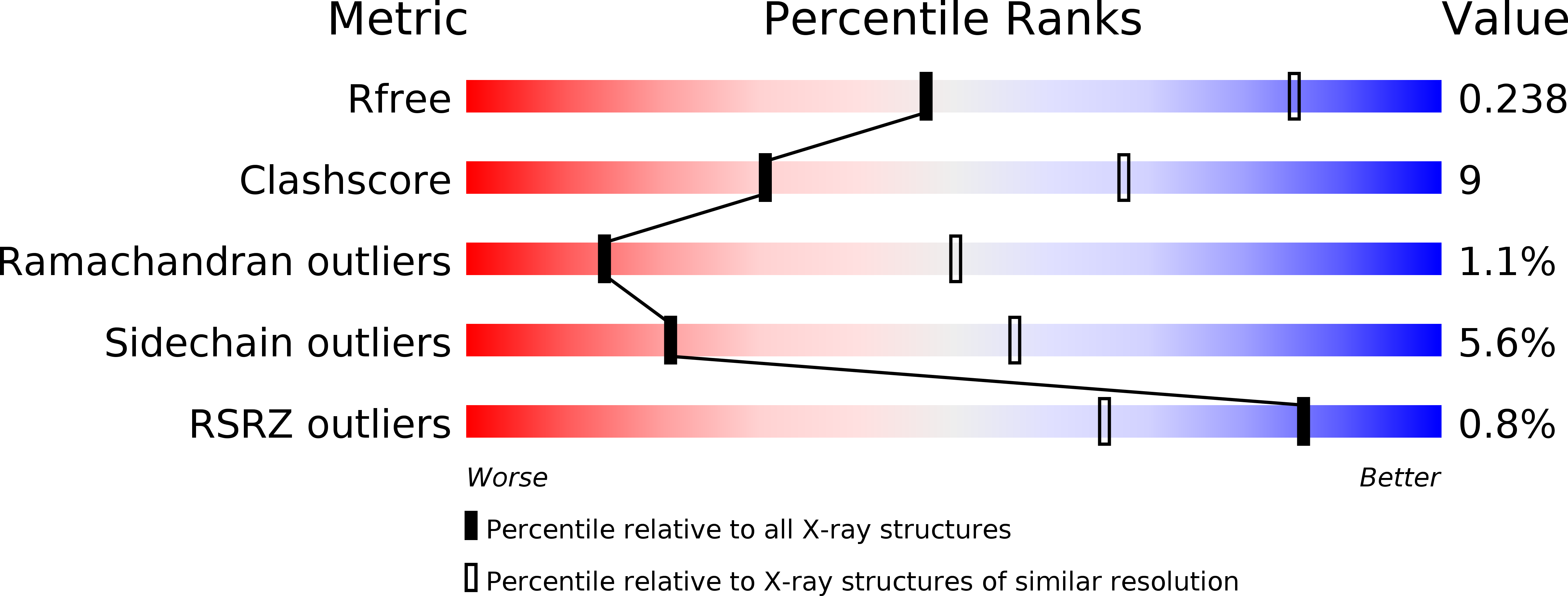
Resolution:
3.00 Å
R-Value Free:
0.25
R-Value Work:
0.20
R-Value Observed:
0.20
Space Group:
P 61
Macromolecular Entities
Polymer Type:polypeptide(L)
Description:26S proteasome regulatory complex subunit p42B
Chain IDs:A
Chain Length:394
Number of Molecules:1
Biological Source:Drosophila melanogaster
Primary Citation
The proteasomal subunit Rpn6 is a molecular clamp holding the core and regulatory subcomplexes together.
Proc.Natl.Acad.Sci.USA 109 149 154 (2012)
PMID: 22187461 DOI: 10.1073/pnas.1117648108

Abstact

Proteasomes execute the degradation of most cellular proteins. Although the 20S core particle (CP) has been studied in great detail, the structure of the 19S regulatory particle (RP), which prepares ubiquitylated substrates for degradation, has remained elusive. Here, we report the crystal structure of one of the RP subunits, Rpn6, and we describe its integration into the cryo-EM density map of the 26S holocomplex at 9.1 Å resolution. Rpn6 consists of an α-solenoid-like fold and a proteasome COP9/signalosome eIF3 (PCI) module in a right-handed suprahelical configuration. Highly conserved surface areas of Rpn6 interact with the conserved surfaces of the Pre8 (alpha2) and Rpt6 subunits from the alpha and ATPase rings, respectively. The structure suggests that Rpn6 has a pivotal role in stabilizing the otherwise weak interaction between the CP and the RP.

Legend

Protein

Chemical

Disease

Primary Citation of related structures