3TW0 image
Entry Detail
PDB ID:
3TW0
Keywords:
Title:
Structural Analysis of Adhesive Tip pilin, GBS104 from Group B Streptococcus agalactiae
Biological Source:
Host Organism:
PDB Version:
Deposition Date:
2011-09-21
Release Date:
2013-03-27
Method Details:
Experimental Method:
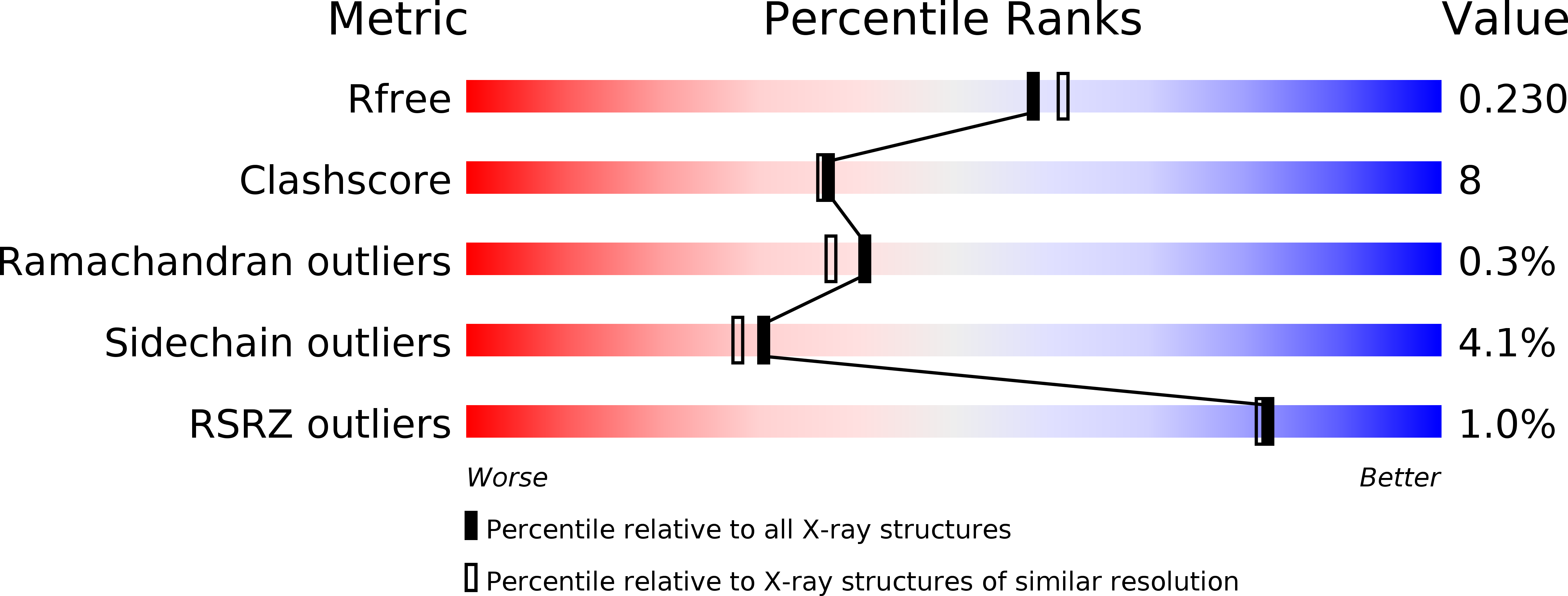
Resolution:
2.00 Å
R-Value Free:
0.23
R-Value Work:
0.19
R-Value Observed:
0.19
Space Group:
P 1
Macromolecular Entities
Polymer Type:polypeptide(L)
Description:Cell wall surface anchor family protein
Mutations:T564C, K571C
Chain IDs:A, B, C, D
Chain Length:370
Number of Molecules:4
Biological Source:Streptococcus agalactiae serogroup V
Primary Citation
Structure of Streptococcus agalactiae tip pilin GBS104: a model for GBS pili assembly and host interactions.
Acta Crystallogr.,Sect.D 69 1073 1089 (2013)
PMID: 23695252 DOI: 10.1107/S0907444913004642

Abstact

The crystal structure of a 75 kDa central fragment of GBS104, a tip pilin from the 2063V/R strain of Streptococcus agalactiae (group B streptococcus; GBS), is reported. In addition, a homology model of the remaining two domains of GBS104 was built and a model of full-length GBS104 was generated by combining the homology model (the N1 and N4 domains) and the crystal structure of the 75 kDa fragment (the N2 and N3 domains). This rod-shaped GBS104 model is constructed of three IgG-like domains (the N1, N2 and N4 domains) and one vWFA-like domain (the N3 domain). The N1 and N2 domains of GBS104 are assembled with distinct and remote segments contributed by the N- and C-termini. The metal-binding site in the N3 domain of GBS104 is in the closed/low-affinity conformation. Interestingly, this domain hosts two long arms that project away from the metal-binding site. Using site-directed mutagenesis, two cysteine residues that lock the N3 domain of GBS104 into the open/high-affinity conformation were introduced. Both wild-type and disulfide-locked recombinant proteins were tested for binding to extracellular matrix proteins such as collagen, fibronectin, fibrinogen and laminin, and an increase in fibronectin binding affinity was identified for the disulfide-locked N3 domain, suggesting that induced conformational changes may play a possible role in receptor binding.

Legend

Protein

Chemical

Disease

Primary Citation of related structures