3TUR image
Deposition Date 2011-09-17
Release Date 2012-12-05
Last Version Date 2024-02-28
Entry Detail
PDB ID:
3TUR
Title:
Crystal Structure of M. tuberculosis LD-transpeptidase type 2 complexed with a peptidoglycan fragment
Biological Source:
Source Organism:
Host Organism:
Method Details:
Experimental Method:
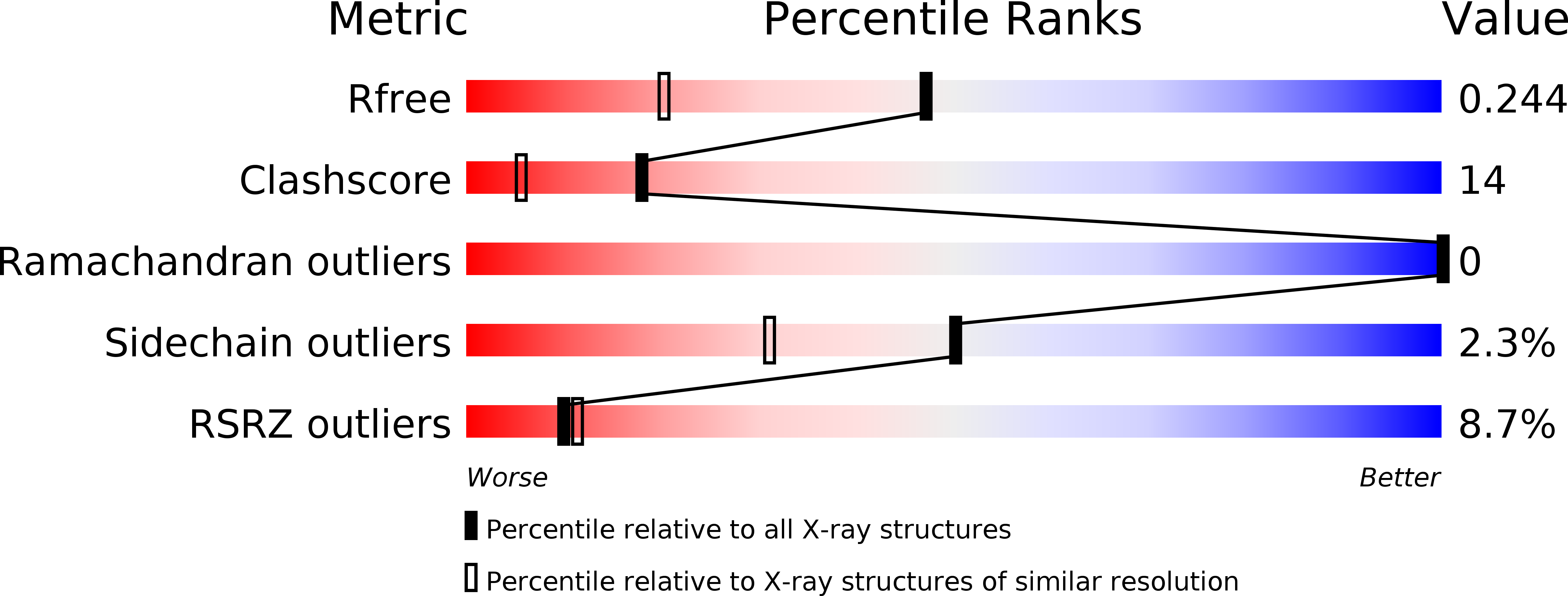
Resolution:
1.72 Å
R-Value Free:
0.23
R-Value Work:
0.19
R-Value Observed:
0.20
Space Group:
I 21 21 21
Macromolecular Entities
Polymer Type:polypeptide(L)
Molecule:Mycobacteria Tuberculosis LD-transpeptidase type 2
Gene (Uniprot):ldtB
Chain IDs:A, B
Chain Length:287
Number of Molecules:2
Biological Source:Mycobacterium tuberculosis
Primary Citation
Targeting the Cell Wall of Mycobacterium tuberculosis: Structure and Mechanism of L,D-Transpeptidase 2.
Structure 20 2103 2115 (2012)
PMID: 23103390 DOI: 10.1016/j.str.2012.09.016

Abstact

With multidrug-resistant cases of tuberculosis increasing globally, better antibiotic drugs and novel drug targets are becoming an urgent need. Traditional β-lactam antibiotics that inhibit D,D-transpeptidases are not effective against mycobacteria, in part because mycobacteria rely mostly on L,D-transpeptidases for biosynthesis and maintenance of their peptidoglycan layer. This reliance plays a major role in drug resistance and persistence of Mycobacterium tuberculosis (Mtb) infections. The crystal structure at 1.7 Å resolution of the Mtb L,D-transpeptidase Ldt(Mt2) containing a bound peptidoglycan fragment, reported here, provides information about catalytic site organization as well as substrate recognition by the enzyme. Based on our structural, kinetic, and calorimetric data, we propose a catalytic mechanism for Ldt(Mt2) in which both acyl-acceptor and acyl-donor substrates reach the catalytic site from the same, rather than different, entrances. Together, this information provides vital insights to facilitate development of drugs targeting this validated yet unexploited enzyme.

Legend

Protein

Chemical

Disease

Primary Citation of related structures