3TUD image
Entry Detail
PDB ID:
3TUD
Title:
Crystal structure of SYK kinase domain with N-(4-methyl-3-(8-methyl-7-oxo-2-(phenylamino)-7,8-dihydropyrido[2,3-d]pyrimidin-6-yl)phenyl)-3-(trifluoromethyl)benzamide
Biological Source:
Source Organism:
PDB Version:
Deposition Date:
2011-09-16
Release Date:
2012-08-29
Method Details:
Experimental Method:
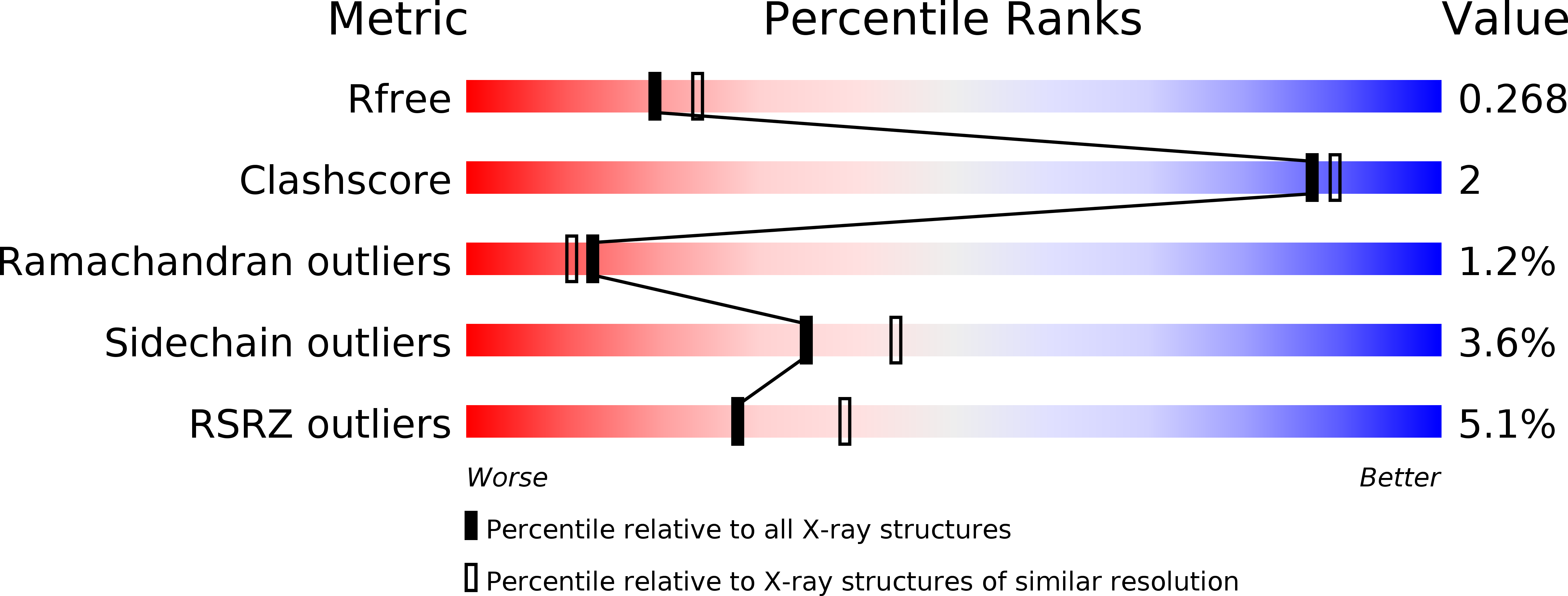
Resolution:
2.33 Å
R-Value Free:
0.26
R-Value Work:
0.19
R-Value Observed:
0.19
Space Group:
P 21 21 21
Macromolecular Entities
Polymer Type:polypeptide(L)
Description:Tyrosine-protein kinase SYK
Chain IDs:A
Chain Length:299
Number of Molecules:1
Biological Source:Homo sapiens
Ligand Molecules
Primary Citation
Identification of Type-II Inhibitors Using Kinase Structures.
Chem.Biol.Drug Des. 80 657 664 (2012)
PMID: 22759374 DOI: 10.1111/j.1747-0285.2012.01443.x

Abstact

Spleen tyrosine kinase is a non-receptor tyrosine kinase, overactivation of which is thought to contribute to autoimmune diseases as well as allergy and asthma. Protein kinases have a highly conserved ATP binding site, thus making challenging the design of selective small molecule inhibitors. It has been well documented that some protein kinases can be stabilized in their inactive conformations (Type-II inhibitors). Herein, we describe a protein structure/ligand-based approach to successfully identify ligands that bind to novel conformations of spleen tyrosine kinase. By utilizing kinase protein crystal structures both in the public domain (RCSB) and within Pfizer's protein crystal database, we report the discovery of the first spleen tyrosine kinase Type-II ligands. Compounds 1 and 3 were found to bind to the DFG-out conformation of spleen tyrosine kinase, while compound 2 binds to a DFG-in, C-Helix-out conformation. In this instance, the C-helix moved significantly to create a large hydrophobic pocket rarely seen in kinase protein crystal structures.

Legend

Protein

Chemical

Disease

Primary Citation of related structures