3TU5 image
Deposition Date 2011-09-15
Release Date 2012-09-26
Last Version Date 2024-02-28
Entry Detail
PDB ID:
3TU5
Title:
Actin complex with Gelsolin Segment 1 fused to Cobl segment
Biological Source:
Source Organism:
Homo sapiens (Taxon ID: 9606)
Mus musculus (Taxon ID: 10090)
Oryctolagus cuniculus (Taxon ID: 9986)
Host Organism:
Method Details:
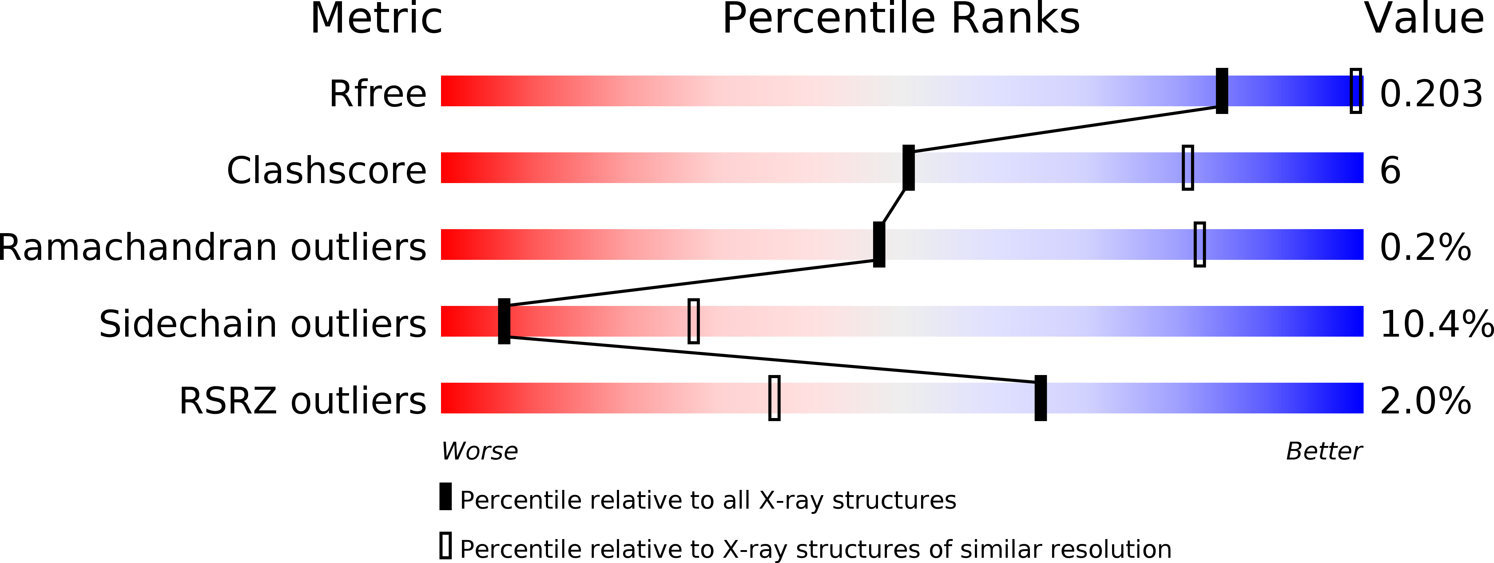
Experimental Method:
Resolution:
3.00 Å
R-Value Free:
0.20
R-Value Work:
0.16
R-Value Observed:
0.16
Space Group:
P 32 2 1
Macromolecular Entities
Polymer Type:polypeptide(L)
Molecule:Actin, alpha skeletal muscle
Gene (Uniprot):ACTA1
Chain IDs:A
Chain Length:377
Number of Molecules:1
Biological Source:Oryctolagus cuniculus
Polymer Type:polypeptide(L)
Molecule:Gelsolin,Protein cordon-bleu,Thymosin beta-4
Gene (Uniprot):GSN, TMSB4X, Cobl
Chain IDs:B
Chain Length:297
Number of Molecules:1
Biological Source:Homo sapiens, Mus musculus
Primary Citation
Structural States and dynamics of the d-loop in actin.
Biophys.J. 103 930 939 (2012)
PMID: 23009842 DOI: 10.1016/j.bpj.2012.07.030

Abstact

Conformational changes induced by ATP hydrolysis on actin are involved in the regulation of complex actin networks. Previous structural and biochemical data implicate the DNase I binding loop (D-loop) of actin in such nucleotide-dependent changes. Here, we investigated the structural and conformational states of the D-loop (in solution) using cysteine scanning mutagenesis and site-directed labeling. The reactivity of D-loop cysteine mutants toward acrylodan and the mobility of spin labels on these mutants do not show patterns of an α-helical structure in monomeric and filamentous actin, irrespective of the bound nucleotide. Upon transition from monomeric to filamentous actin, acrylodan emission spectra and electron paramagnetic resonance line shapes of labeled mutants are blue-shifted and more immobilized, respectively, with the central residues (residues 43-47) showing the most drastic changes. Moreover, complex electron paramagnetic resonance line shapes of spin-labeled mutants suggest several conformational states of the D-loop. Together with a new (to our knowledge) actin crystal structure that reveals the D-loop in a unique hairpin conformation, our data suggest that the D-loop equilibrates in F-actin among different conformational states irrespective of the nucleotide state of actin.

Legend

Protein

Chemical

Disease

Primary Citation of related structures