Deposition Date
2011-09-14
Release Date
2011-10-05
Last Version Date
2024-11-27
Entry Detail
PDB ID:
3TTB
Keywords:
Title:
Structure of the Thioalkalivibrio paradoxus cytochrome c nitrite reductase in complex with sulfite
Biological Source:
Source Organism(s):
Thioalkalivibrio paradoxus (Taxon ID: 108010)
Method Details:
Experimental Method:
Resolution:
2.00 Å
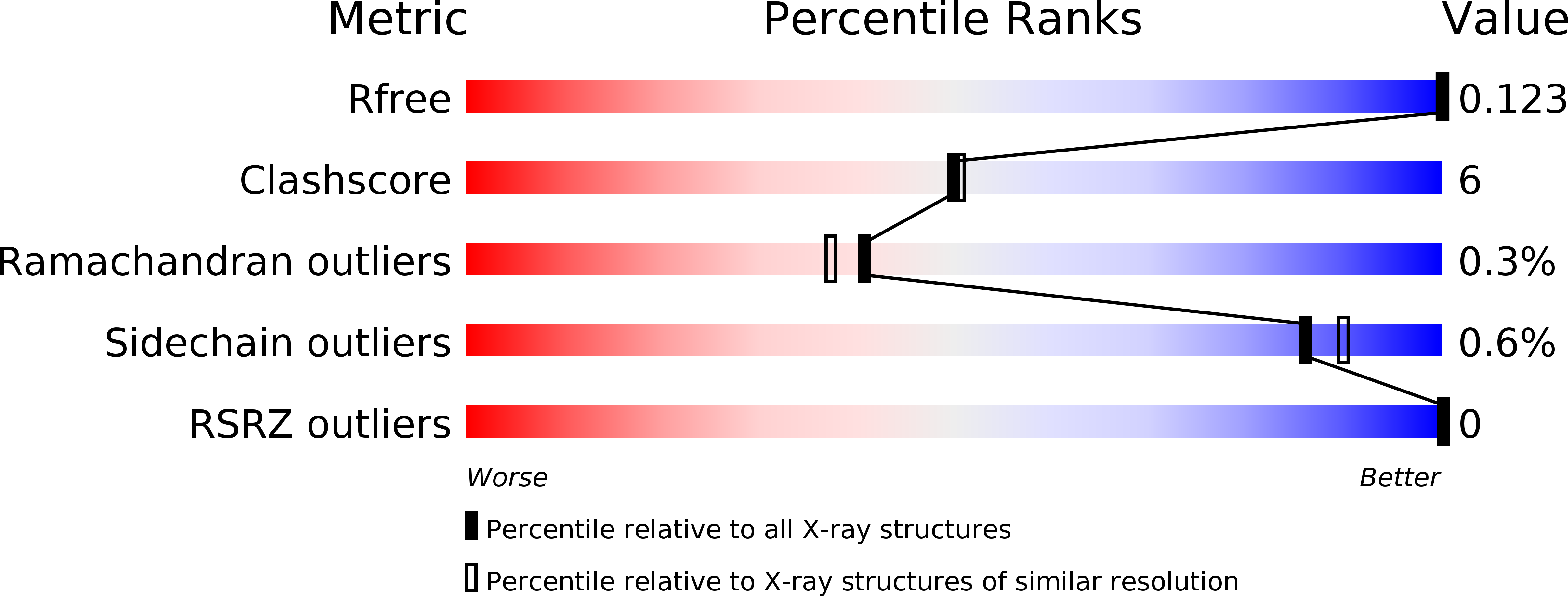
R-Value Free:
0.15
R-Value Work:
0.13
R-Value Observed:
0.13
Space Group:
P 21 3