Deposition Date
2011-09-12
Release Date
2012-02-22
Last Version Date
2024-02-28
Entry Detail
PDB ID:
3TS9
Keywords:
Title:
Crystal Structure of the MDA5 Helicase Insert Domain
Biological Source:
Source Organism(s):
Mus musculus (Taxon ID: 10090)
Expression System(s):
Method Details:
Experimental Method:
Resolution:
2.00 Å
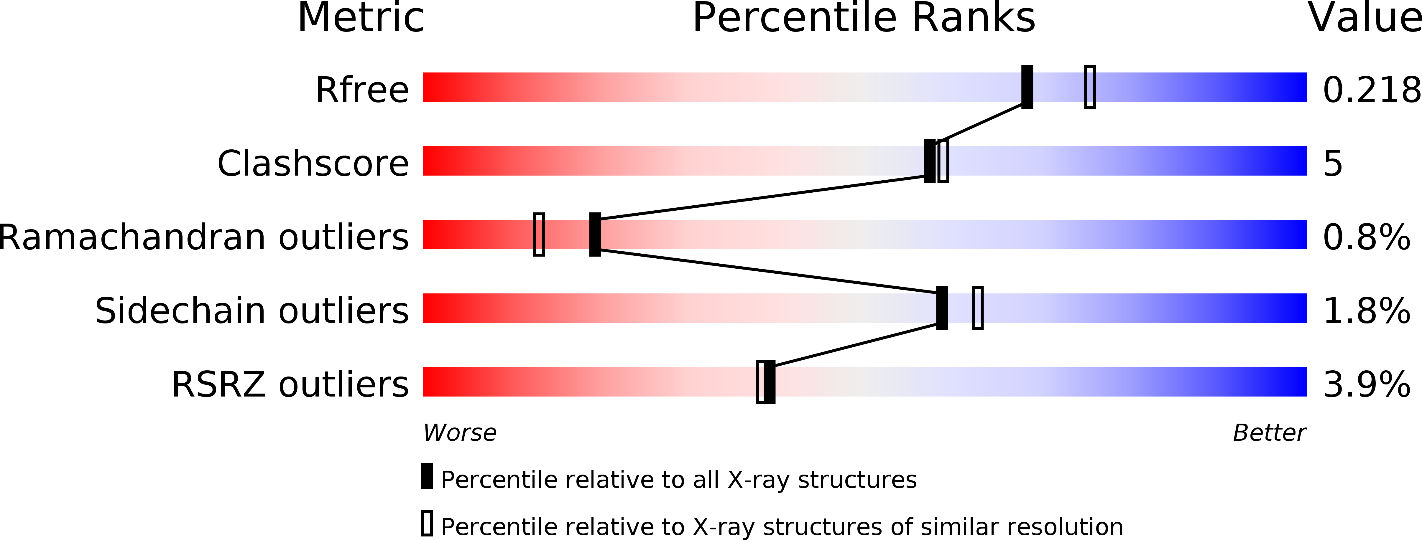
R-Value Free:
0.22
R-Value Work:
0.19
R-Value Observed:
0.19
Space Group:
P 21 21 21