Deposition Date
2011-09-08
Release Date
2012-06-20
Last Version Date
2023-12-06
Entry Detail
PDB ID:
3TPX
Keywords:
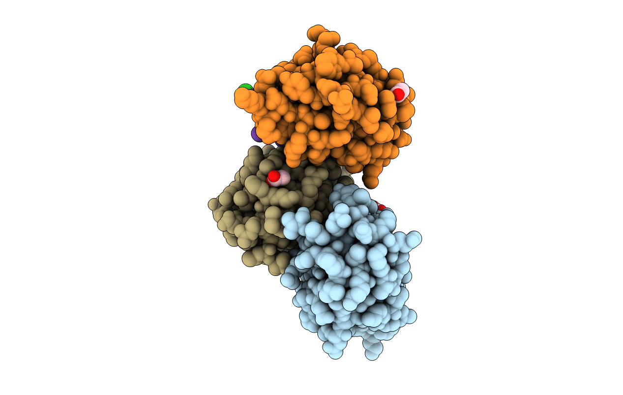
Title:
Crystal structure of human MDM2 in complex with a trifluoromethylated D-peptide inhibitor
Biological Source:
Source Organism(s):
Homo sapiens (Taxon ID: 9606)
Method Details:
Experimental Method:
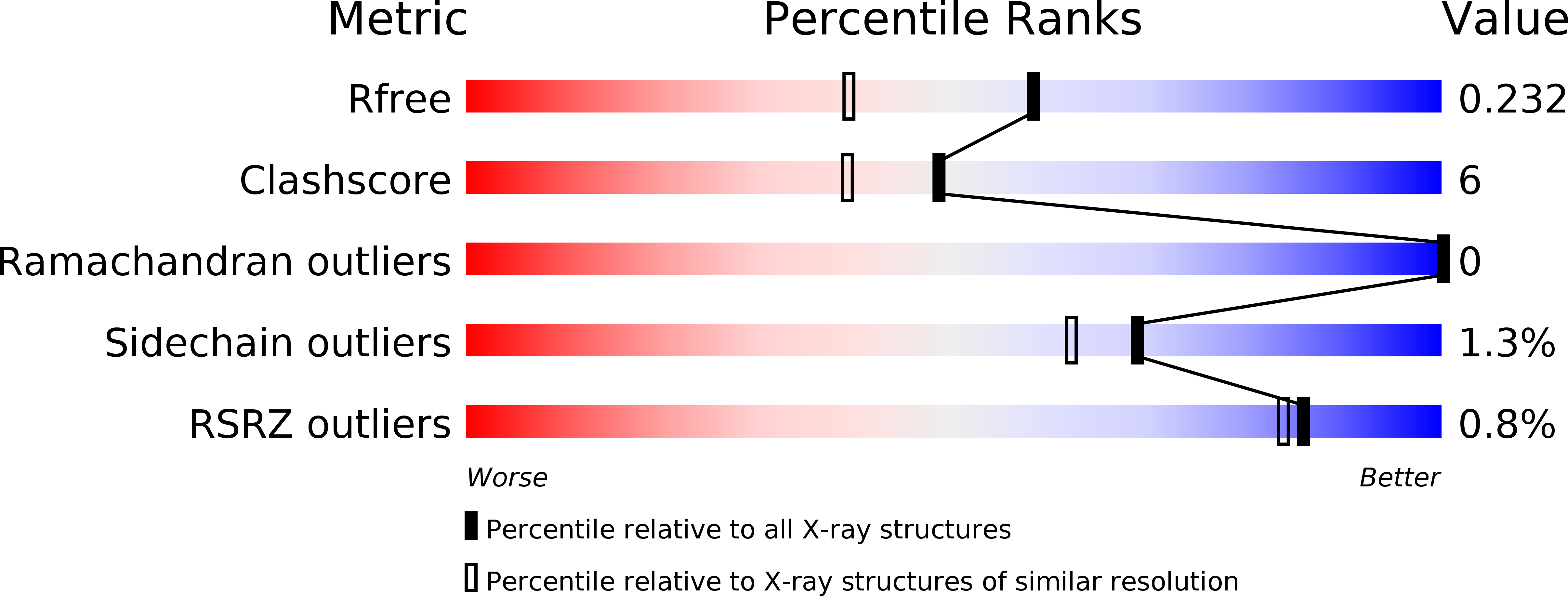
Resolution:
1.80 Å
R-Value Free:
0.23
R-Value Work:
0.19
R-Value Observed:
0.19
Space Group:
C 2 2 21