3TNQ image
Deposition Date 2011-09-01
Release Date 2012-02-01
Last Version Date 2024-11-20
Entry Detail
PDB ID:
3TNQ
Keywords:
Title:
Structure and Allostery of the PKA RIIb Tetrameric Holoenzyme
Biological Source:
Source Organism:
Rattus norvegicus (Taxon ID: 10116)
Mus musculus (Taxon ID: 10090)
Host Organism:
Method Details:
Experimental Method:
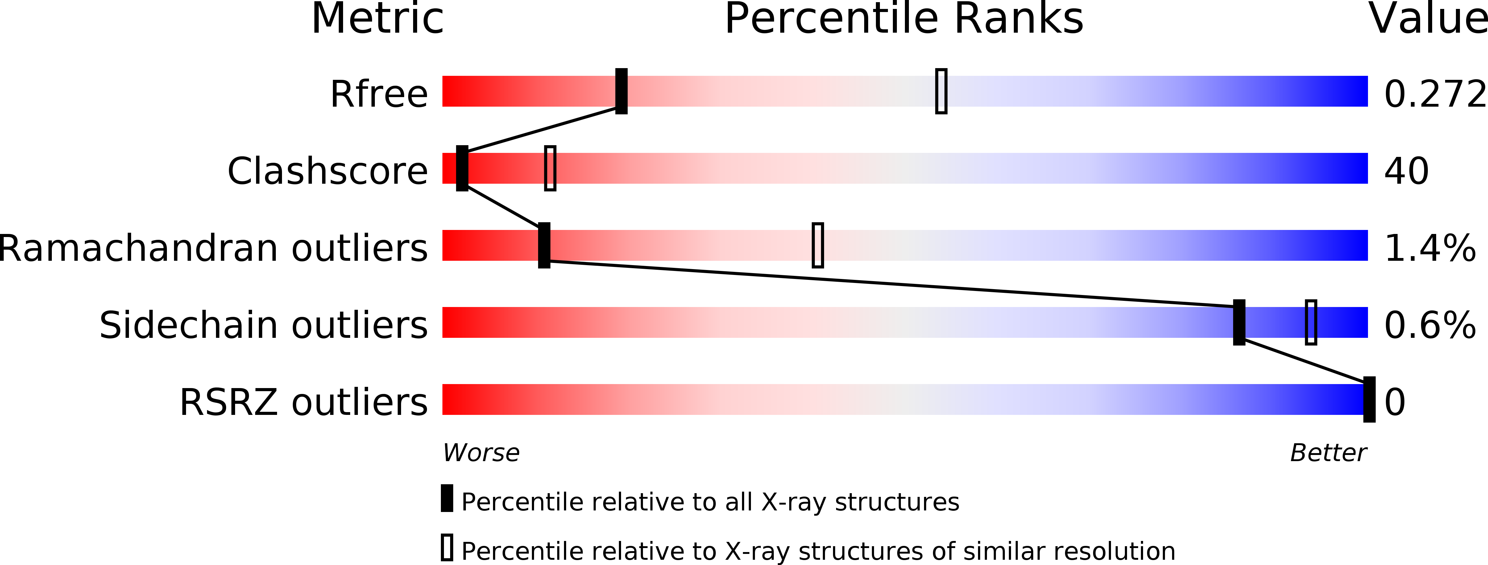
Resolution:
3.10 Å
R-Value Free:
0.27
R-Value Work:
0.23
R-Value Observed:
0.23
Space Group:
C 2 2 2
Macromolecular Entities
Structures with similar UniProt ID
Protein Blast
Polymer Type:polypeptide(L)
Molecule:cAMP-dependent protein kinase type II-beta regulatory subunit
Gene (Uniprot):Prkar2b
Mutagens:R230K
Chain IDs:B (auth: A)
Chain Length:416
Number of Molecules:1
Biological Source:Mus musculus
Structures with similar UniProt ID
Protein Blast
Polymer Type:polypeptide(L)
Molecule:Protein kinase, cAMP-dependent, catalytic, alpha
Chain IDs:A (auth: B)
Chain Length:350
Number of Molecules:1
Biological Source:Rattus norvegicus
Modified Residue
Compound ID Chain ID Parent Comp ID Details 2D Image
SEP A SER PHOSPHOSERINE
TPO A THR PHOSPHOTHREONINE
Primary Citation
Structure and allostery of the PKA RIIbeta tetrameric holoenzyme
Science 335 712 716 (2012)
PMID: 22323819 DOI: 10.1126/science.1213979

Abstact

In its physiological state, cyclic adenosine monophosphate (cAMP)-dependent protein kinase (PKA) is a tetramer that contains a regulatory (R) subunit dimer and two catalytic (C) subunits. We describe here the 2.3 angstrom structure of full-length tetrameric RIIβ(2):C(2) holoenzyme. This structure showing a dimer of dimers provides a mechanistic understanding of allosteric activation by cAMP. The heterodimers are anchored together by an interface created by the β4-β5 loop in the RIIβ subunit, which docks onto the carboxyl-terminal tail of the adjacent C subunit, thereby forcing the C subunit into a fully closed conformation in the absence of nucleotide. Diffusion of magnesium adenosine triphosphate (ATP) into these crystals trapped not ATP, but the reaction products, adenosine diphosphate and the phosphorylated RIIβ subunit. This complex has implications for the dissociation-reassociation cycling of PKA. The quaternary structure of the RIIβ tetramer differs appreciably from our model of the RIα tetramer, confirming the small-angle x-ray scattering prediction that the structures of each PKA tetramer are different.

Legend

Protein

Chemical

Disease

Primary Citation of related structures