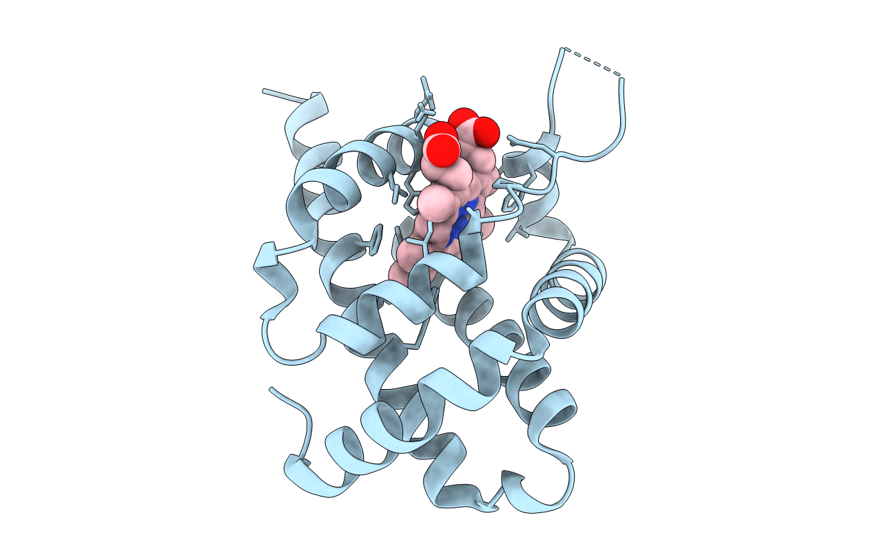
3TM3 image
Entry Detail
PDB ID:
3TM3
Title:
Wild-type hemoglobin from Vitreoscilla stercoraria
Biological Source:
Source Organism:
Host Organism:
PDB Version:
Deposition Date:
2011-08-31
Release Date:
2014-04-16
Method Details:
Experimental Method:
Resolution:
1.75 Å
R-Value Free:
0.20
R-Value Work:
0.17
R-Value Observed:
0.17
Space Group:
C 2 2 21
Macromolecular Entities
Polymer Type:polypeptide(L)
Description:Hemoglobin
Chain IDs:A
Chain Length:146
Number of Molecules:1
Biological Source:Vitreoscilla stercoraria
Ligand Molecules
Primary Citation
Crystallographic structure determination of B10 mutants of Vitreoscilla hemoglobin: role of Tyr29 (B10) in the structure of the ligand-binding site.
Acta Crystallogr.,Sect.F 69 215 222 (2013)
PMID: 23519792 DOI: 10.1107/S1744309112044818

Abstact

Site-directed mutants of the gene encoding wild-type Vitreoscilla hemoglobin were made that changed Tyr29 (B10) of the wild-type Vitreoscilla hemoglobin (VHb) to either Phe or Ala. The wild-type and the two mutant hemoglobins were expressed in Escherichia coli and purified to homogeneity. The binding of the two mutants to CO was essentially identical to that of wild-type VHb as determined by CO-difference spectra. Circular-dichroism spectra also showed the two mutants to be essentially the same as wild-type VHb regarding overall helicity. All three VHbs were crystallized and their structures were determined at resolutions of 1.7-1.9 Å, which are similar to that of the original wild-type structure determination. The Tyr29Phe mutant has a structure that is essentially indistinguishable from that of the wild type. However, the structure of the Tyr29Ala mutant has significant differences from that of the wild type. In addition, for the Tyr29Ala mutant it was possible to determine the positions of most of the residues in the D region, which was disordered in the originally reported structure of wild-type VHb as well as in the wild-type VHb structure reported here. In the Tyr29Ala mutant, the five-membered ring of proline E8 (Pro54) occupies the space occupied by the aromatic ring of Tyr29 in the wild-type structure. These results are discussed in the context of the proposed role of Tyr29 in the structure of the oxygen-binding pocket.

Legend

Protein

Chemical

Disease

Primary Citation of related structures