3TIP image
Entry Detail
PDB ID:
3TIP
Title:
Crystal structure of Staphylococcus aureus SasG E-G52 module
Biological Source:
Host Organism:
PDB Version:
Deposition Date:
2011-08-21
Release Date:
2012-04-18
Method Details:
Experimental Method:
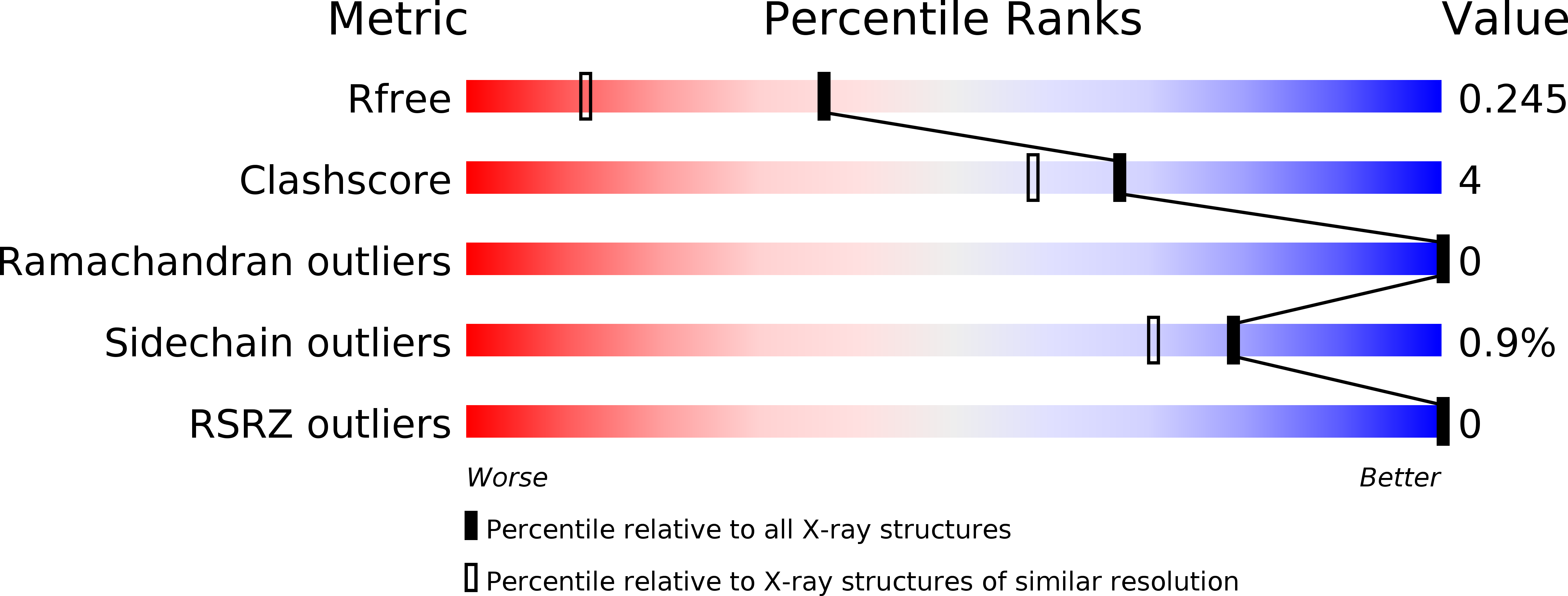
Resolution:
1.70 Å
R-Value Free:
0.23
R-Value Work:
0.19
R-Value Observed:
0.19
Space Group:
P 1 21 1
Macromolecular Entities
Polymer Type:polypeptide(L)
Description:Surface protein G
Chain IDs:A
Chain Length:132
Number of Molecules:1
Biological Source:Staphylococcus aureus subsp. aureus
Primary Citation
Staphylococcal biofilm-forming protein has a contiguous rod-like structure.
Proc.Natl.Acad.Sci.USA 109 E1011 E1018 (2012)
PMID: 22493247 DOI: 10.1073/pnas.1119456109

Abstact

Staphylococcus aureus and Staphylococcus epidermidis form communities (called biofilms) on inserted medical devices, leading to infections that affect many millions of patients worldwide and cause substantial morbidity and mortality. As biofilms are resistant to antibiotics, device removal is often required to resolve the infection. Thus, there is a need for new therapeutic strategies and molecular data that might assist their development. Surface proteins S. aureus surface protein G (SasG) and accumulation-associated protein (S. epidermidis) promote biofilm formation through their "B" regions. B regions contain tandemly arrayed G5 domains interspersed with approximately 50 residue sequences (herein called E) and have been proposed to mediate intercellular accumulation through Zn(2+)-mediated homodimerization. Although E regions are predicted to be unstructured, SasG and accumulation-associated protein form extended fibrils on the bacterial surface. Here we report structures of E-G5 and G5-E-G5 from SasG and biophysical characteristics of single and multidomain fragments. E sequences fold cooperatively and form interlocking interfaces with G5 domains in a head-to-tail fashion, resulting in a contiguous, elongated, monomeric structure. E and G5 domains lack a compact hydrophobic core, and yet G5 domain and multidomain constructs have thermodynamic stabilities only slightly lower than globular proteins of similar size. Zn(2+) does not cause SasG domains to form dimers. The work reveals a paradigm for formation of fibrils on the 100-nm scale and suggests that biofilm accumulation occurs through a mechanism distinct from the "zinc zipper." Finally, formation of two domains by each repeat (as in SasG) might reduce misfolding in proteins when the tandem arrangement of highly similar sequences is advantageous.

Legend

Protein

Chemical

Disease

Primary Citation of related structures