3THK image
Deposition Date 2011-08-19
Release Date 2011-11-23
Last Version Date 2023-09-13
Entry Detail
PDB ID:
3THK
Title:
Structure of SH3 chimera with a type II ligand linked to the chain C-terminal
Biological Source:
Source Organism:
Host Organism:
Method Details:
Experimental Method:
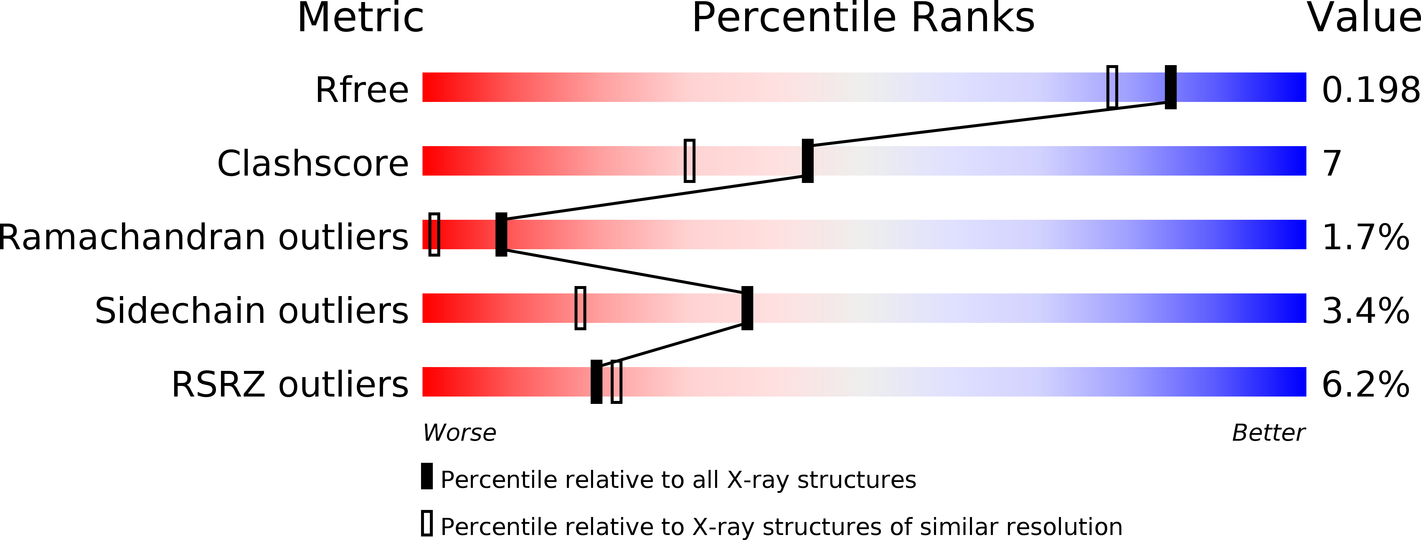
Resolution:
1.70 Å
R-Value Free:
0.20
R-Value Work:
0.17
R-Value Observed:
0.18
Space Group:
P 32
Macromolecular Entities
Structures with similar UniProt ID
Protein Blast
Polymer Type:polypeptide(L)
Molecule:Spectrin alpha chain, brain
Gene (Uniprot):Sptan1
Chain IDs:A, B
Chain Length:73
Number of Molecules:2
Biological Source:Rattus norvegicus
Polymer Type:polypeptide(L)
Molecule:Proline-rich peptide
Chain IDs:C, D
Chain Length:10
Number of Molecules:2
Biological Source:
Primary Citation
High-Resolution Crystal Structure of Spectrin SH3 Domain Fused with a Proline-Rich Peptide.
J.Biomol.Struct.Dyn. 29 485 495 (2011)
PMID: 22066535

Abstact

A new chimeric protein, named WT-CIIA, was designed by connecting the proline-rich decapeptide PPPVPPYSAG to the C-terminus of the alpha-spectrin SH3 domain through a natural twelve-residue linker to obtain a single-chain model that would imitate intramolecular SH3-ligand interaction. The crystal structure of this fusion protein was determined at 1.7 Å resolution. The asymmetric unit of the crystal contained two SH3 globules contacting with one PPPVPPY fragment located between them. The domains are related by the two-fold non-crystallographic axis and the ligand lies in two opposite orientations with respect to the conservative binding sites of SH3 domains.

Legend

Protein

Chemical

Disease

Primary Citation of related structures