Deposition Date
2011-08-17
Release Date
2012-07-04
Last Version Date
2023-09-13
Entry Detail
PDB ID:
3TGU
Keywords:
Title:
Cytochrome bc1 complex from chicken with pfvs-designed moa inhibitor bound
Biological Source:
Source Organism(s):
Gallus gallus (Taxon ID: 9031)
Method Details:
Experimental Method:
Resolution:
2.70 Å
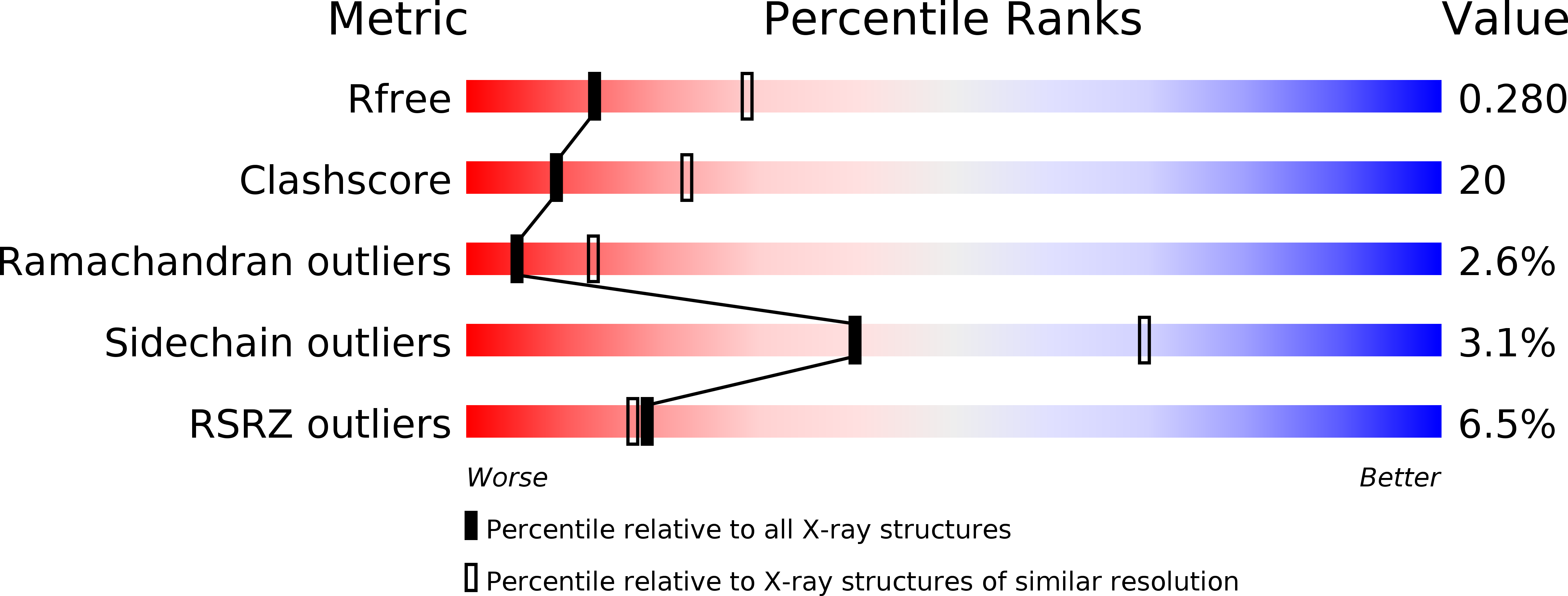
R-Value Free:
0.28
R-Value Work:
0.25
R-Value Observed:
0.25
Space Group:
P 21 21 21