3TGC image
Deposition Date 2011-08-17
Release Date 2012-08-22
Last Version Date 2024-10-16
Entry Detail
PDB ID:
3TGC
Title:
Crystal structure of L130R mutant of Nitrophorin 4 from Rhodnius prolixus complexed with nitrite at pH 7.4
Biological Source:
Source Organism:
Host Organism:
Method Details:
Experimental Method:
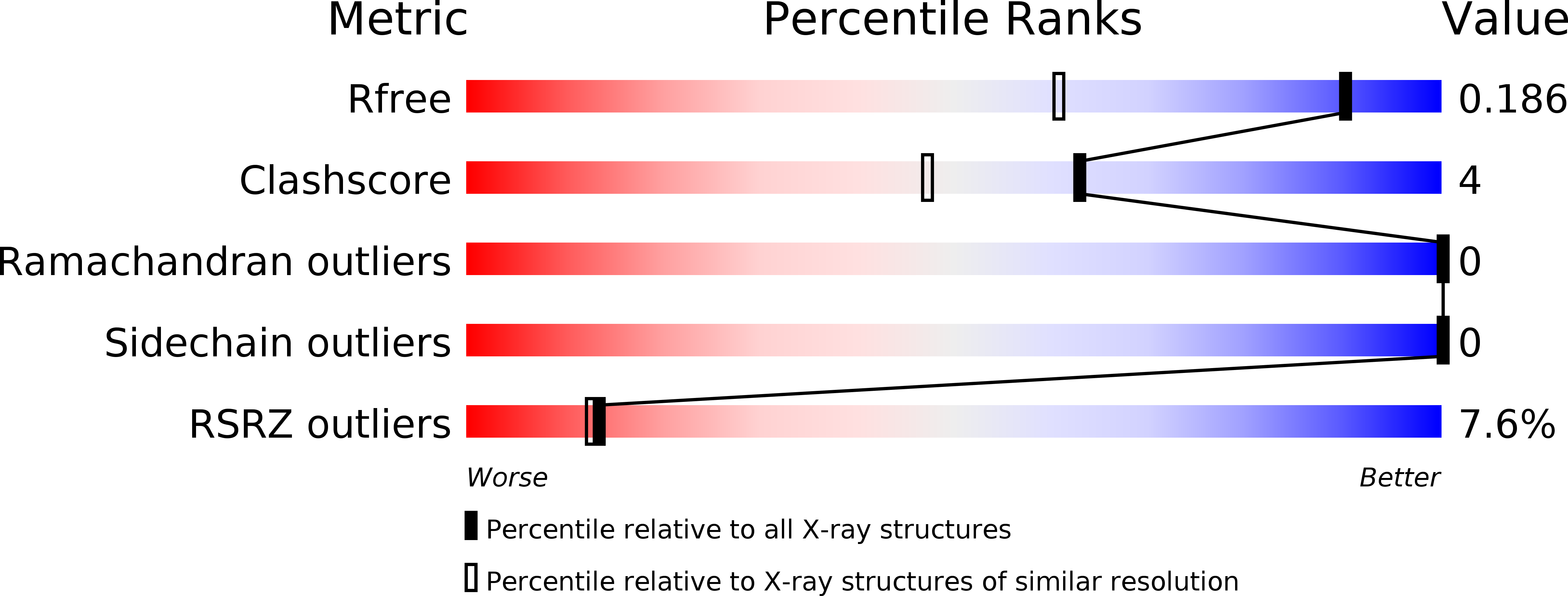
Resolution:
1.40 Å
R-Value Free:
0.18
R-Value Work:
0.14
R-Value Observed:
0.15
Space Group:
C 1 2 1
Macromolecular Entities
Structures with similar UniProt ID
Protein Blast
Polymer Type:polypeptide(L)
Molecule:Nitrophorin-4
Mutagens:L130R
Chain IDs:A
Chain Length:184
Number of Molecules:1
Biological Source:Rhodnius prolixus
Primary Citation
Insertion of an H-Bonding Residue into the Distal Pocket of the Ferriheme Protein Nitrophorin 4: Effect on Nitrite[BOND]Iron Coordination and Nitrite Disproportionation
Chem.Biodivers. 9 1761 1775 (2012)
PMID: 22976968 DOI: 10.1002/cbdv.201100401

Abstact

Heme proteins are important entities for the metabolism of nitrite. Inspection of the structural features of the reported hemoprotein-nitrite crystal structures reveals that, except for nitrophorin 4 (NP4), H-bonding to the nitrite ligand is accomplished via histidine or arginine residues. These H-bonds probably play an important role for the nitrite coordination and/or reactivities. In nitrophorins, which catalyze the nitrite disproportionation reaction, such a residue is missing. Here, we report on the L130R mutant of the NP isoprotein NP4 that provides the Arg130 residue as part of the flexible G-H loop as a potential H-bonding residue in the distal heme pocket. Similar to the wild-type protein, nitrite remains N-bonded in the crystal structure of NP4(L130R). However, spectroscopic investigations show that, in solution, a second ligand-rotational orientation exists, which is in fast-exchange equilibrium with the normal, parallel ligand orientation. Moreover, the nitrite disproportionation is inhibited in NP4(L130R). Comparison with another, also less active mutant NP4(D30N) suggests that the displacement of H(2)O molecules from the heme cavity prevents the proton donation pathway through Asp30.

Legend

Protein

Chemical

Disease

Primary Citation of related structures