Deposition Date
2011-08-12
Release Date
2011-09-21
Last Version Date
2023-09-13
Entry Detail
PDB ID:
3TE7
Keywords:
Title:
Quinone Oxidoreductase (NQ02) bound to the imidazoacridin-6-one 5a1
Biological Source:
Source Organism(s):
Homo sapiens (Taxon ID: 9606)
Expression System(s):
Method Details:
Experimental Method:
Resolution:
1.70 Å
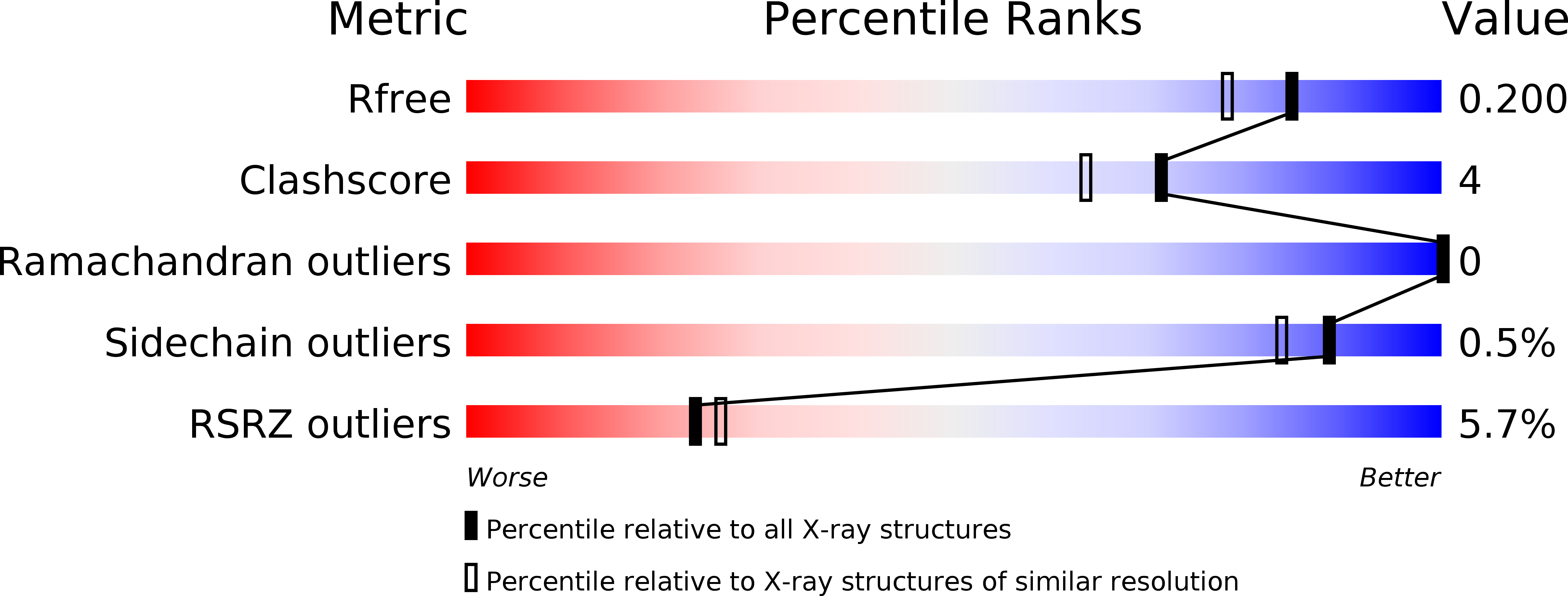
R-Value Free:
0.20
R-Value Work:
0.16
R-Value Observed:
0.17
Space Group:
P 21 21 21