Deposition Date
2011-08-09
Release Date
2012-10-03
Last Version Date
2025-10-22
Entry Detail
PDB ID:
3TCQ
Keywords:
Title:
Crystal Structure of matrix protein VP40 from Ebola virus Sudan
Biological Source:
Source Organism(s):
Sudan ebolavirus (Taxon ID: 128948)
Expression System(s):
Method Details:
Experimental Method:
Resolution:
1.60 Å
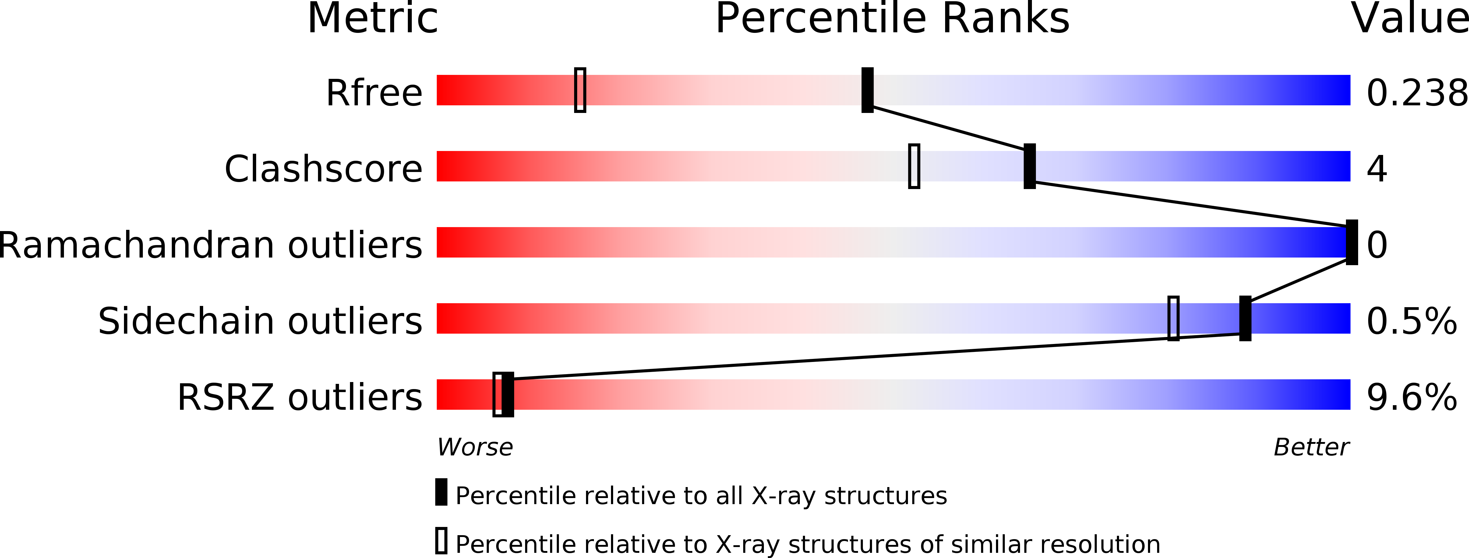
R-Value Free:
0.22
R-Value Work:
0.19
R-Value Observed:
0.19
Space Group:
C 1 2 1