Planned Maintenance: Some services may turn out to be unavailable from 15th January, 2026 to 16th January, 2026. We apologize for the inconvenience!

3TBN image
Deposition Date 2011-08-07
Release Date 2011-10-05
Last Version Date 2024-03-20
Entry Detail
PDB ID:
3TBN
Title:
Crystal structure of a miner2 homolog: a type 6 CDGSH iron-sulfur protein.
Biological Source:
Source Organism(s):
Expression System(s):
Method Details:
Experimental Method:
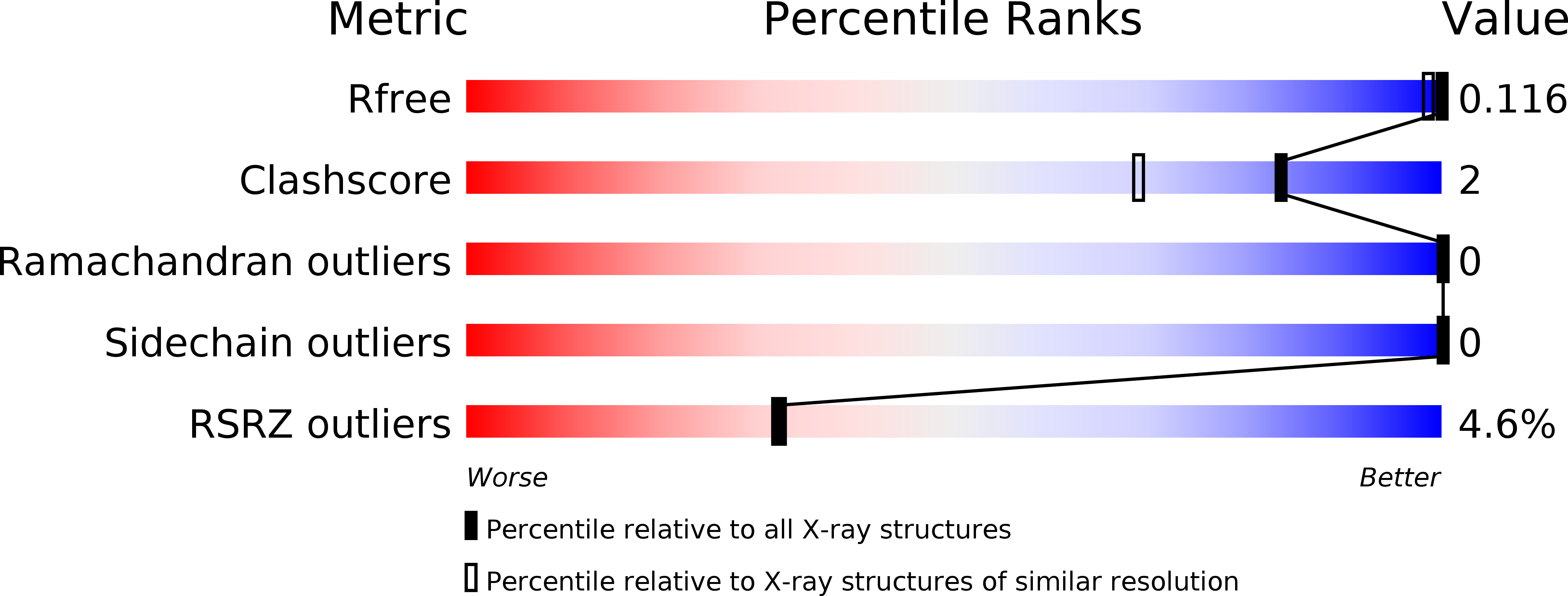
Resolution:
1.15 Å
R-Value Free:
0.12
R-Value Work:
0.11
R-Value Observed:
0.11
Space Group:
P 65
Macromolecular Entities
Structures with similar UniProt ID
Protein Blast
Polymer Type:polypeptide(L)
Molecule:Putative uncharacterized protein
Gene (Uniprot):amb4007
Chain IDs:A
Chain Length:87
Number of Molecules:1
Biological Source:Magnetospirillum magneticum
Ligand Molecules
Primary Citation
Structure and Molecular Evolution of CDGSH Iron-Sulfur Domains.
Plos One 6 e24790 e24790 (2011)
PMID: 21949752 DOI: 10.1371/journal.pone.0024790

Abstact

The recently discovered CDGSH iron-sulfur domains (CISDs) are classified into seven major types with a wide distribution throughout the three domains of life. The type 1 protein mitoNEET has been shown to fold into a dimer with the signature CDGSH motif binding to a [2Fe-2S] cluster. However, the structures of all other types of CISDs were unknown. Here we report the crystal structures of type 3, 4, and 6 CISDs determined at 1.5 Å, 1.8 Å and 1.15 Å resolution, respectively. The type 3 and 4 CISD each contain one CDGSH motif and adopt a dimeric structure. Although similar to each other, the two structures have permutated topologies, and both are distinct from the type 1 structure. The type 6 CISD contains tandem CDGSH motifs and adopts a monomeric structure with an internal pseudo dyad symmetry. All currently known CISD structures share dual iron-sulfur binding modules and a β-sandwich for either intermolecular or intramolecular dimerization. The iron-sulfur binding module, the β-strand N-terminal to the module and a proline motif are conserved among different type structures, but the dimerization module and the interface and orientation between the two iron-sulfur binding modules are divergent. Sequence analysis further shows resemblance between CISD types 4 and 7 and between 1 and 2. Our findings suggest that all CISDs share common ancestry and diverged into three primary folds with a characteristic phylogenetic distribution: a eukaryote-specific fold adopted by types 1 and 2 proteins, a prokaryote-specific fold adopted by types 3, 4 and 7 proteins, and a tandem-motif fold adopted by types 5 and 6 proteins. Our comprehensive structural, sequential and phylogenetic analysis provides significant insight into the assembly principles and evolutionary relationship of CISDs.

Legend

Protein

Chemical

Disease

Primary Citation of related structures
Feedback Form
Name
Email
Institute
Feedback