Deposition Date
2011-08-07
Release Date
2011-10-05
Last Version Date
2024-03-20
Entry Detail
PDB ID:
3TBM
Keywords:
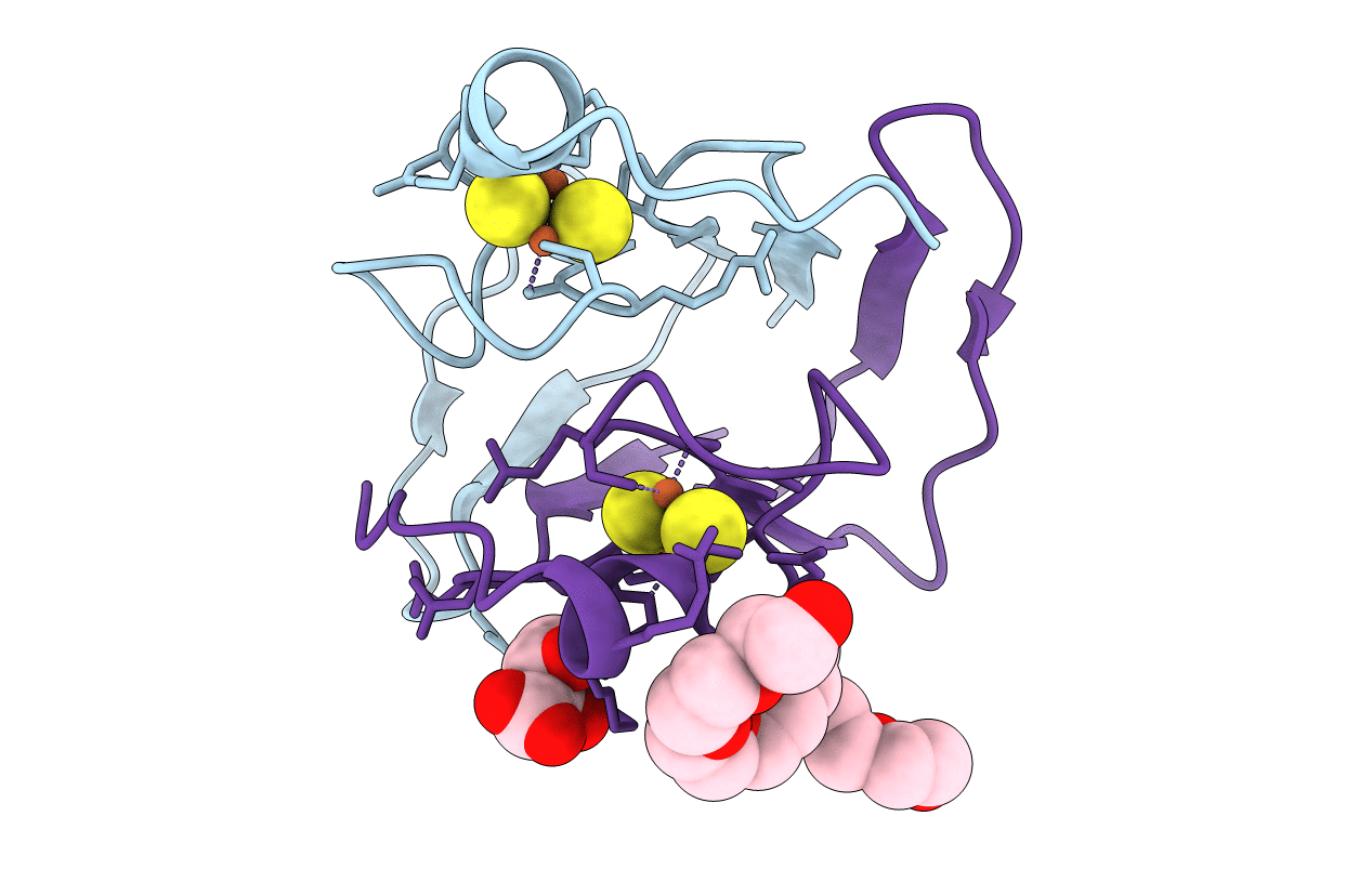
Title:
Crystal structure of a type 4 CDGSH iron-sulfur protein.
Biological Source:
Source Organism(s):
Ralstonia solanacearum (Taxon ID: 267608)
Expression System(s):
Method Details:
Experimental Method:
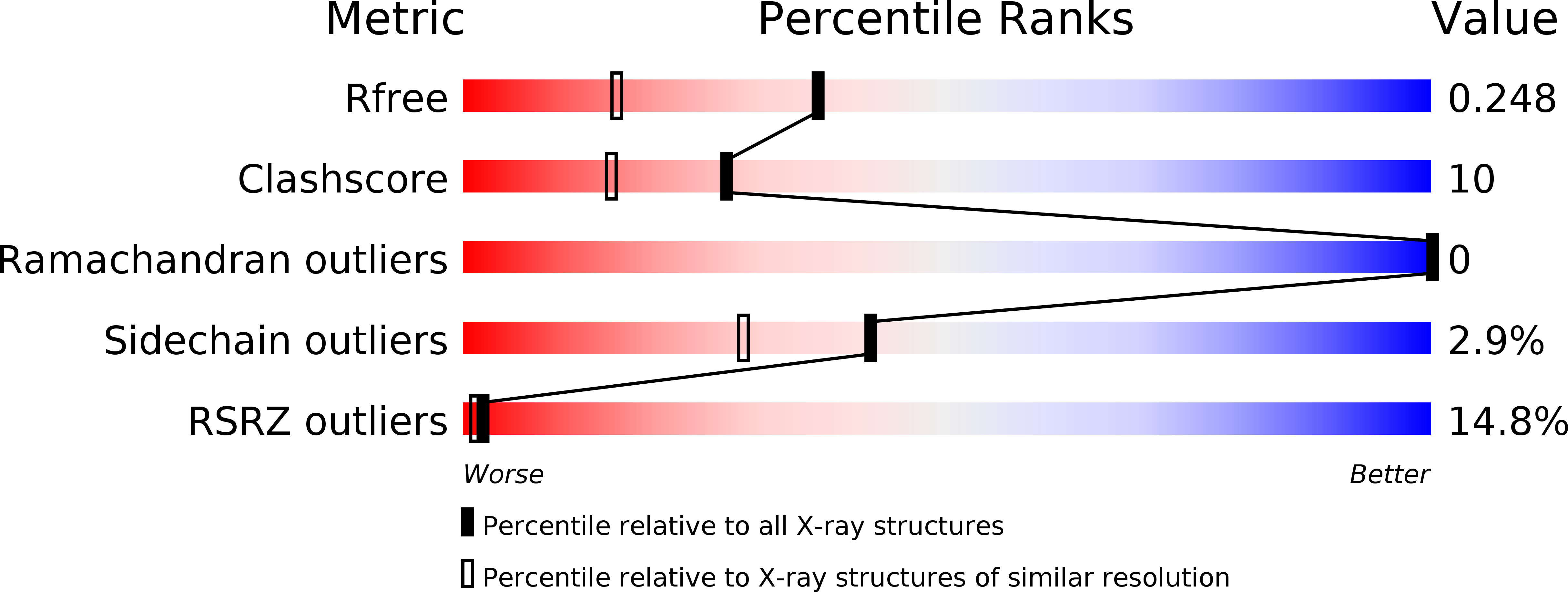
Resolution:
1.80 Å
R-Value Free:
0.25
R-Value Work:
0.20
R-Value Observed:
0.20
Space Group:
C 1 2 1