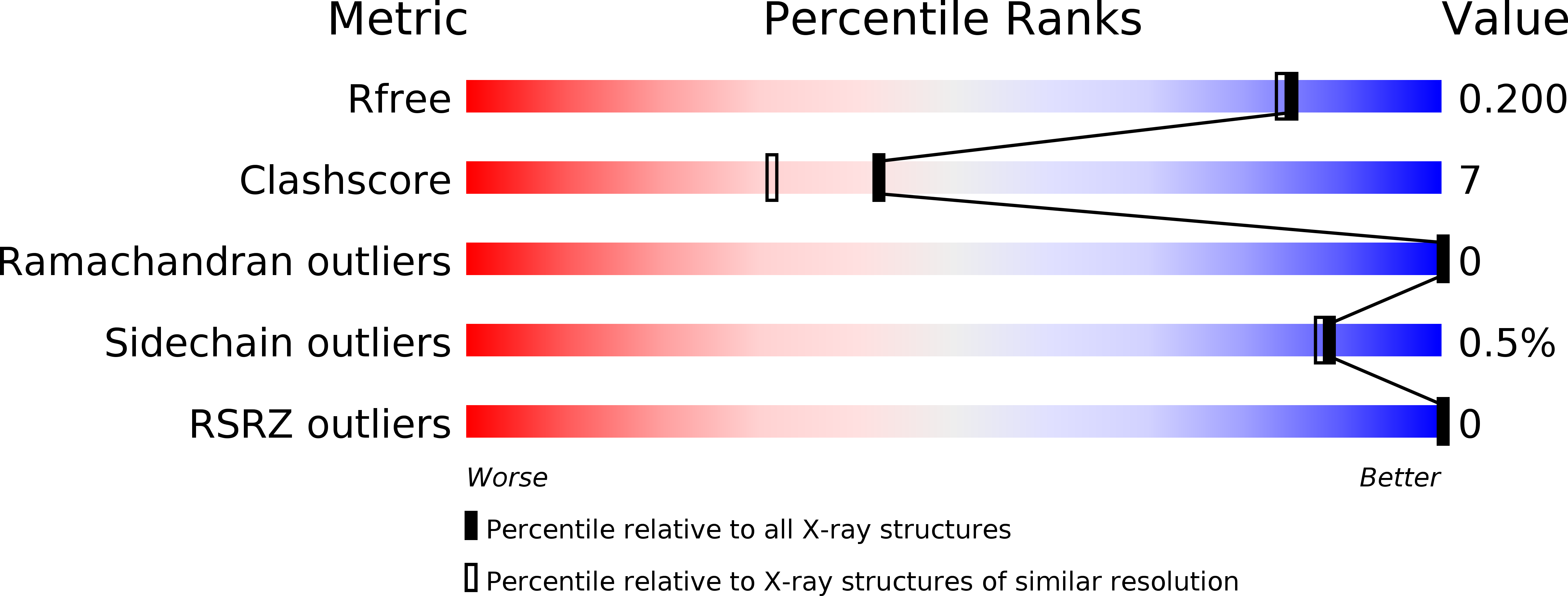
3TBJ image
Deposition Date 2011-08-07
Release Date 2012-08-22
Last Version Date 2024-11-27
Entry Detail
PDB ID:
3TBJ
Keywords:
Title:
The 1.7A crystal structure of Actibind a T2 ribonucleases as antitumorigenic agents
Biological Source:
Source Organism:
Method Details:
Experimental Method:
Resolution:
1.80 Å
R-Value Free:
0.18
R-Value Work:
0.15
R-Value Observed:
0.15
Space Group:
P 32 2 1
Macromolecular Entities
Polymer Type:polypeptide(L)
Molecule:Actibind
Gene (Uniprot):ACTIB-I
Chain IDs:A
Chain Length:237
Number of Molecules:1
Biological Source:Aspergillus niger
Modified Residue
Compound ID Chain ID Parent Comp ID Details 2D Image
ASN A ASN GLYCOSYLATION SITE
Primary Citation
The 1.8 A crystal structure of ACTIBIND suggests a mode of action for T2 ribonucleases as antitumorigenic agents.
J.Med.Chem. 55 1013 1020 (2012)
PMID: 22216760 DOI: 10.1021/jm1015507

Abstact

ACTIBIND and its human homologue RNASET2 are T2 ribonucleases (RNases). RNases are ubiquitous and efficient enzymes that hydrolyze RNA to 3' mononucleotides and also possess antitumorigenic and antiangiogenic activities. Previously, we have shown that ACTIBIND and RNASET2 bind actin and interfere with the cytoskeletal network structure, thereby inhibiting cell motility and invasiveness in cancer and in endothelial cells. We also showed that ACTIBIND binds actin in a molar ratio of 1:2. Here, we further characterize ACTIBIND and determine its crystal structure at 1.8 Å resolution, which enables us to propose two structural elements that create binding sites to actin. We suggest that each of these binding sites is composed of one cysteine residue and one conserved amino acid region. These binding sites possibly interfere with the cytoskeleton network structure and as such may be responsible for the antitumorigenic and antiangiogenic activities of ACTIBIND and its human analogue RNASET2.

Legend

Protein

Chemical

Disease

Primary Citation of related structures