3TB6 image
Deposition Date 2011-08-05
Release Date 2012-01-25
Last Version Date 2023-09-13
Entry Detail
PDB ID:
3TB6
Title:
Structure of the effector-binding domain of arabinose repressor AraR from Bacillus subtilis
Biological Source:
Source Organism:
Host Organism:
Method Details:
Experimental Method:
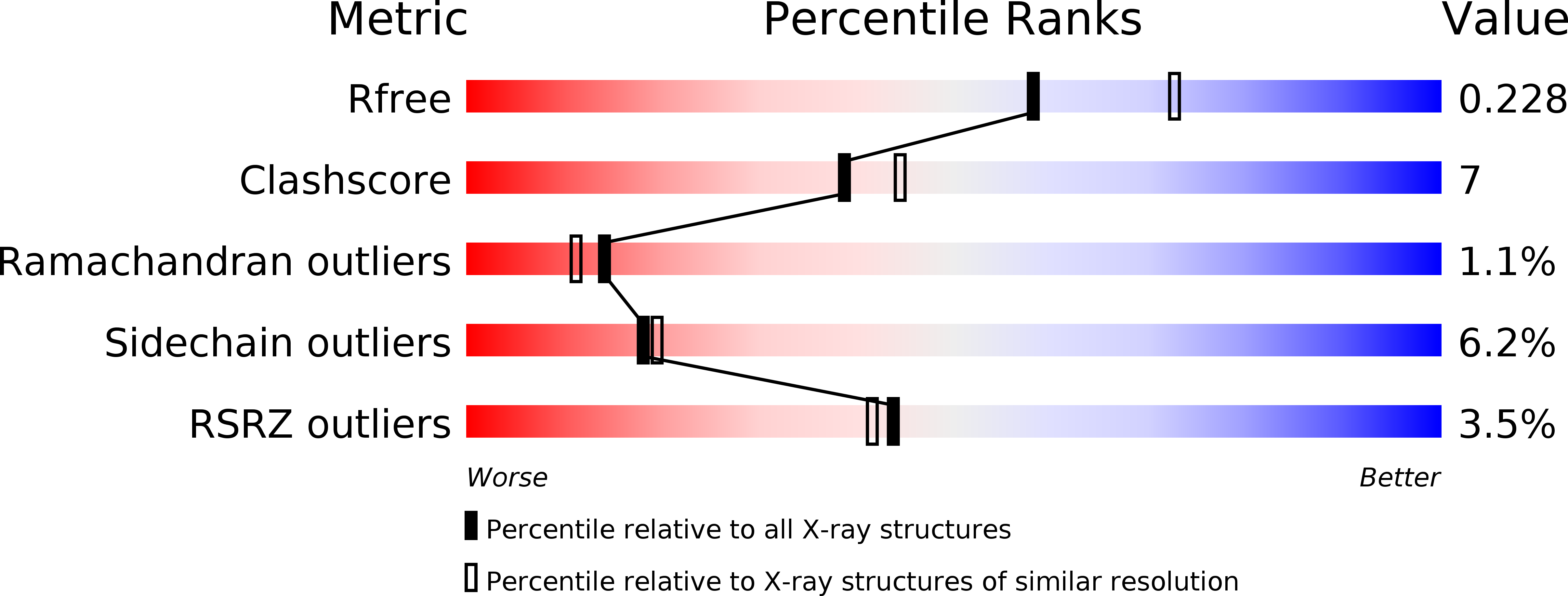
Resolution:
2.21 Å
R-Value Free:
0.23
R-Value Work:
0.18
R-Value Observed:
0.18
Space Group:
P 21 21 21
Macromolecular Entities
Structures with similar UniProt ID
Protein Blast
Polymer Type:polypeptide(L)
Molecule:Arabinose metabolism transcriptional repressor
Gene (Uniprot):araR
Chain IDs:A, B
Chain Length:298
Number of Molecules:2
Biological Source:Bacillus subtilis
Primary Citation
Structure of the effector-binding domain of the arabinose repressor AraR from Bacillus subtilis.
Acta Crystallogr.,Sect.D 68 176 185 (2012)
PMID: 22281747 DOI: 10.1107/S090744491105414X

Abstact

In Bacillus subtilis, the arabinose repressor AraR negatively controls the expression of genes in the metabolic pathway of arabinose-containing polysaccharides. The protein is composed of two domains of different phylogenetic origin and function: an N-terminal DNA-binding domain belonging to the GntR family and a C-terminal effector-binding domain that shows similarity to members of the GalR/LacI family. The crystal structure of the C-terminal effector-binding domain of AraR in complex with the effector L-arabinose has been determined at 2.2 Å resolution. The L-arabinose binding affinity was characterized by isothermal titration calorimetry and differential scanning fluorimetry; the K(d) value was 8.4 ± 0.4 µM. The effect of L-arabinose on the protein oligomeric state was investigated in solution and detailed analysis of the crystal identified a dimer organization which is distinctive from that of other members of the GalR/LacI family.

Legend

Protein

Chemical

Disease

Primary Citation of related structures