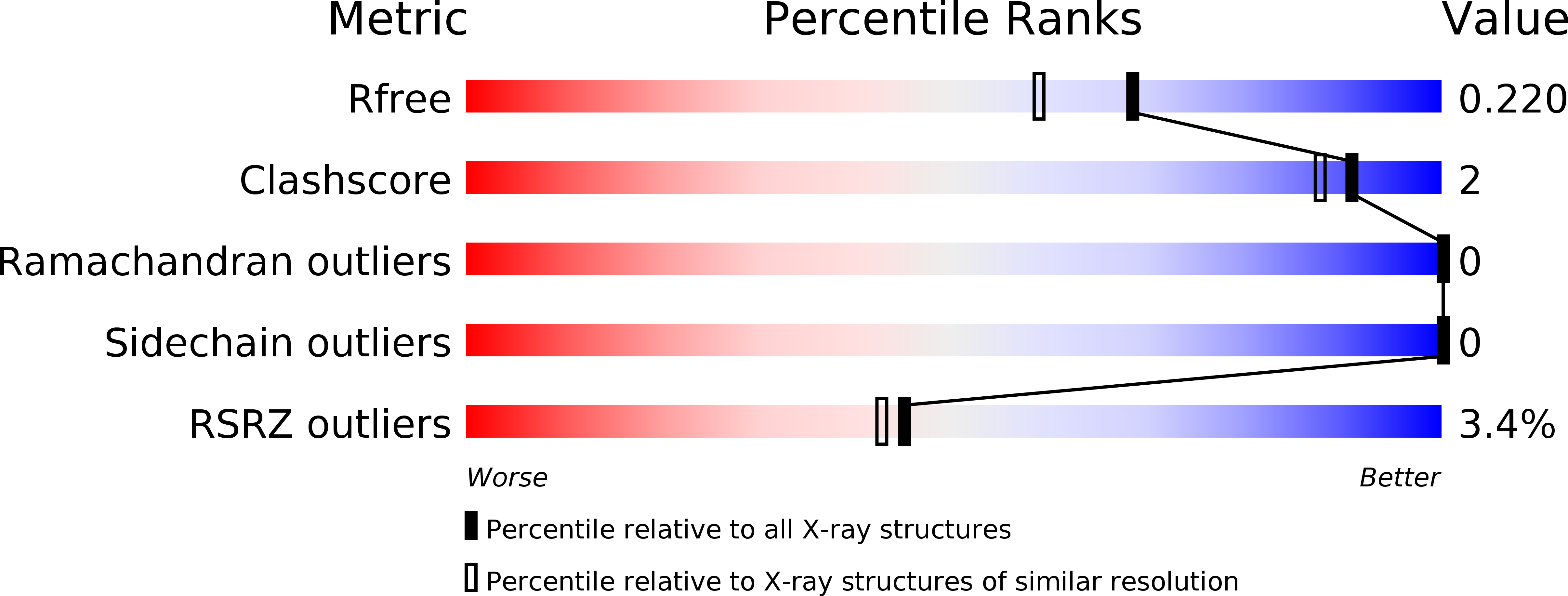
3TAH image
Deposition Date 2011-08-04
Release Date 2011-12-14
Last Version Date 2023-11-01
Entry Detail
PDB ID:
3TAH
Keywords:
Title:
Crystal structure of an S. thermophilus NFeoB N11A mutant bound to mGDP
Biological Source:
Source Organism:
Host Organism:
Method Details:
Experimental Method:
Resolution:
1.85 Å
R-Value Free:
0.21
R-Value Work:
0.17
R-Value Observed:
0.17
Space Group:
C 2 2 21
Macromolecular Entities
Structures with similar UniProt ID
Protein Blast
Polymer Type:polypeptide(L)
Molecule:Ferrous iron uptake transporter protein B
Gene (Uniprot):feoB
Mutagens:N11A
Chain IDs:A, B
Chain Length:272
Number of Molecules:2
Biological Source:Streptococcus thermophilus
Primary Citation
The structure of an N11A mutant of the G-protein domain of FeoB
Acta Crystallogr.,Sect.F 67 1511 1515 (2011)
PMID: 22139154 DOI: 10.1107/S1744309111042965

Abstact

The uptake of ferrous iron in prokaryotes is mediated by the G-protein-coupled membrane protein FeoB. The protein contains two N-terminal soluble domains that are together called `NFeoB'. One of these is a G-protein domain, and GTP hydrolysis by this domain is essential for iron transport. The GTPase activity of NFeoB is accelerated in the presence of potassium ions, which bind at a site adjacent to the nucleotide. One of the ligands at the potassium-binding site is a conserved asparagine residue, which corresponds to Asn11 in Streptococcus thermophilus NFeoB. The structure of an N11A S. thermophilus NFeoB mutant has been determined and refined to a resolution of 1.85 Å; the crystals contained a mixture of mant-GDP-bound and mant-GMP-bound protein. The structure demonstrates how the use of a derivatized nucleotide in cocrystallization experiments can facilitate the growth of diffraction-quality crystals.

Legend

Protein

Chemical

Disease

Primary Citation of related structures