Deposition Date
2011-07-25
Release Date
2011-10-12
Last Version Date
2024-02-28
Entry Detail
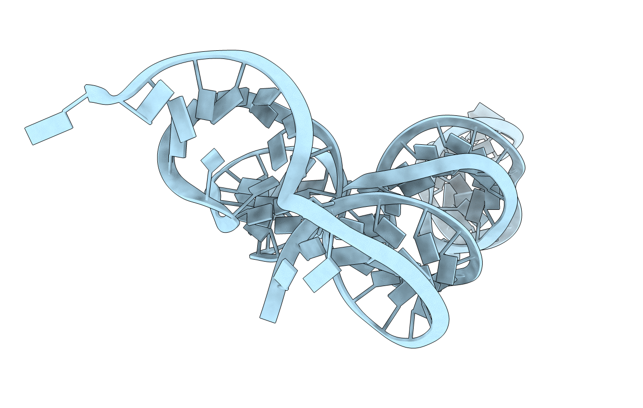
Biological Source:
Source Organism(s):
synthetic construct (Taxon ID: 32630)
Hepatitis C virus ED43 (Taxon ID: 356418)
Hepatitis C virus ED43 (Taxon ID: 356418)
Method Details:
Experimental Method:
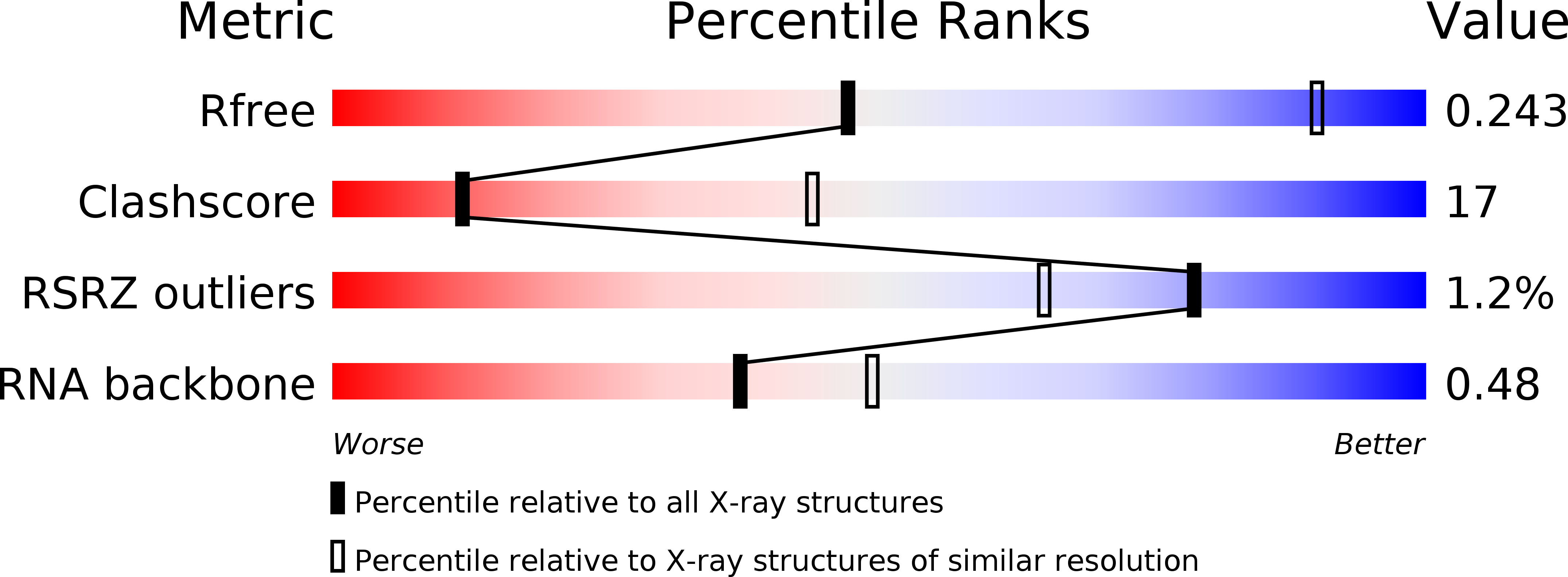
Resolution:
3.55 Å
R-Value Free:
0.25
R-Value Work:
0.22
R-Value Observed:
0.22
Space Group:
P 41 21 2