3T1Y image
Deposition Date 2011-07-22
Release Date 2012-01-25
Last Version Date 2023-09-13
Entry Detail
PDB ID:
3T1Y
Title:
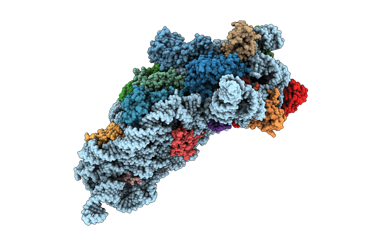
Structure of the Thermus thermophilus 30S ribosomal subunit complexed with a human anti-codon stem loop (HASL) of transfer RNA Lysine 3 (TRNALYS3) bound to an mRNA with an AAG-codon in the A-site and paromomycin
Biological Source:
Source Organism:
Method Details:
Experimental Method:
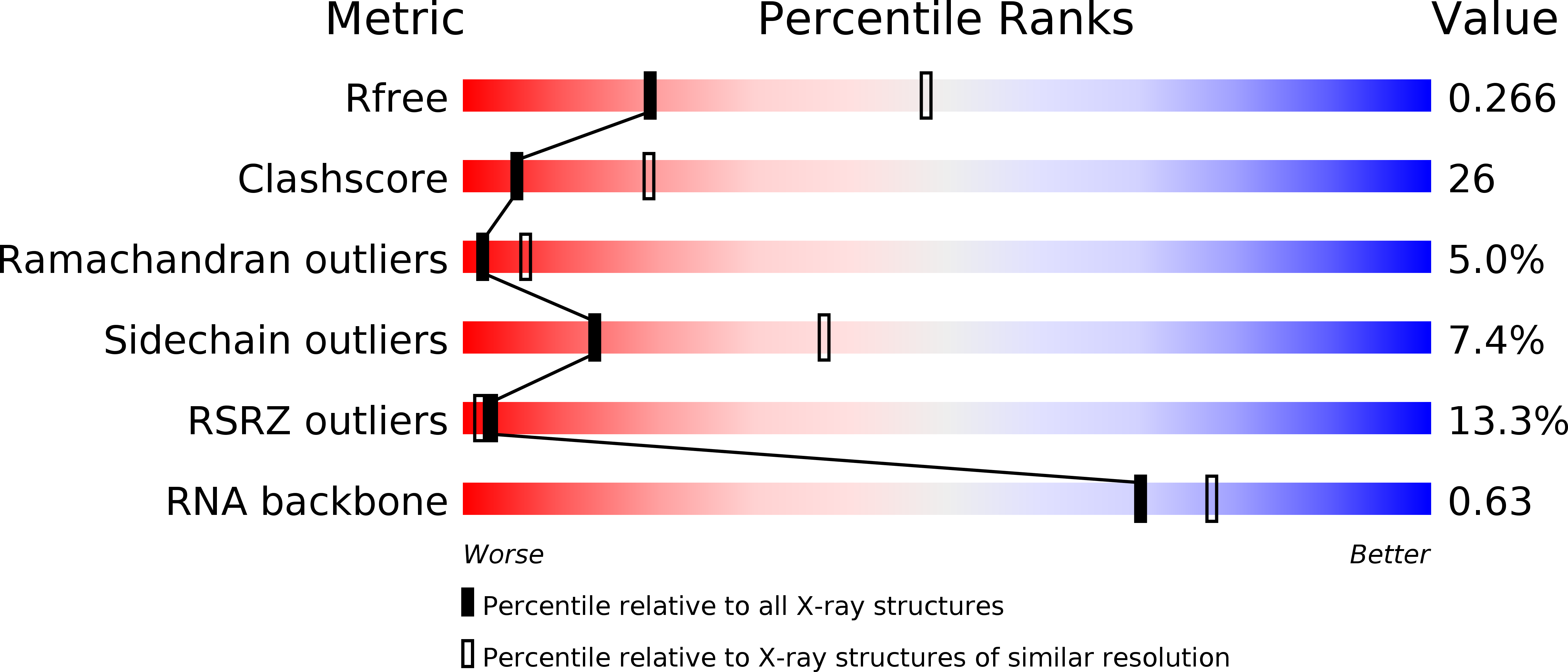
Resolution:
2.80 Å
R-Value Free:
0.26
R-Value Work:
0.24
Space Group:
P 41 21 2
Macromolecular Entities
Polymer Type:polyribonucleotide
Molecule:16S rRNA
Chain IDs:A
Chain Length:1513
Number of Molecules:1
Biological Source:Thermus thermophilus
Structures with similar UniProt ID
Protein Blast
Polymer Type:polypeptide(L)
Molecule:30S ribosomal protein S2
Gene (Uniprot):rpsB
Chain IDs:B
Chain Length:256
Number of Molecules:1
Biological Source:Thermus thermophilus
Structures with similar UniProt ID
Protein Blast
Polymer Type:polypeptide(L)
Molecule:30S ribosomal protein S3
Gene (Uniprot):rpsC
Chain IDs:C
Chain Length:239
Number of Molecules:1
Biological Source:Thermus thermophilus
Structures with similar UniProt ID
Protein Blast
Polymer Type:polypeptide(L)
Molecule:30S ribosomal protein S4
Gene (Uniprot):rpsD
Chain IDs:D
Chain Length:209
Number of Molecules:1
Biological Source:Thermus thermophilus
Structures with similar UniProt ID
Protein Blast
Polymer Type:polypeptide(L)
Molecule:30S ribosomal protein S5
Gene (Uniprot):rpsE
Chain IDs:E
Chain Length:162
Number of Molecules:1
Biological Source:Thermus thermophilus
Structures with similar UniProt ID
Protein Blast
Polymer Type:polypeptide(L)
Molecule:30S ribosomal protein S6
Gene (Uniprot):rpsF
Chain IDs:F
Chain Length:101
Number of Molecules:1
Biological Source:Thermus thermophilus
Structures with similar UniProt ID
Protein Blast
Polymer Type:polypeptide(L)
Molecule:30S ribosomal protein S7
Gene (Uniprot):rpsG
Chain IDs:G
Chain Length:156
Number of Molecules:1
Biological Source:Thermus thermophilus
Structures with similar UniProt ID
Protein Blast
Polymer Type:polypeptide(L)
Molecule:30S ribosomal protein S8
Chain IDs:H
Chain Length:138
Number of Molecules:1
Biological Source:Thermus thermophilus
Structures with similar UniProt ID
Protein Blast
Polymer Type:polypeptide(L)
Molecule:30S ribosomal protein S9
Chain IDs:I
Chain Length:128
Number of Molecules:1
Biological Source:Thermus thermophilus
Structures with similar UniProt ID
Protein Blast
Polymer Type:polypeptide(L)
Molecule:30S ribosomal protein S10
Gene (Uniprot):rpsJ
Chain IDs:J
Chain Length:105
Number of Molecules:1
Biological Source:Thermus thermophilus
Structures with similar UniProt ID
Protein Blast
Polymer Type:polypeptide(L)
Molecule:30S ribosomal protein S11
Gene (Uniprot):rpsK
Chain IDs:K
Chain Length:129
Number of Molecules:1
Biological Source:Thermus thermophilus
Structures with similar UniProt ID
Protein Blast
Polymer Type:polypeptide(L)
Molecule:30S ribosomal protein S12
Gene (Uniprot):rpsL
Chain IDs:L
Chain Length:132
Number of Molecules:1
Biological Source:Thermus thermophilus
Structures with similar UniProt ID
Protein Blast
Polymer Type:polypeptide(L)
Molecule:30S ribosomal protein S13
Gene (Uniprot):rpsM
Chain IDs:M
Chain Length:126
Number of Molecules:1
Biological Source:Thermus thermophilus
Structures with similar UniProt ID
Protein Blast
Polymer Type:polypeptide(L)
Molecule:30S ribosomal protein S14 type Z
Chain IDs:N
Chain Length:61
Number of Molecules:1
Biological Source:Thermus thermophilus
Structures with similar UniProt ID
Protein Blast
Polymer Type:polypeptide(L)
Molecule:30S ribosomal protein S15
Gene (Uniprot):rpsO
Chain IDs:O
Chain Length:89
Number of Molecules:1
Biological Source:Thermus thermophilus
Structures with similar UniProt ID
Protein Blast
Polymer Type:polypeptide(L)
Molecule:30S ribosomal protein S16
Gene (Uniprot):rpsP
Chain IDs:P
Chain Length:88
Number of Molecules:1
Biological Source:Thermus thermophilus
Structures with similar UniProt ID
Protein Blast
Polymer Type:polypeptide(L)
Molecule:30S ribosomal protein S17
Chain IDs:Q
Chain Length:105
Number of Molecules:1
Biological Source:Thermus thermophilus
Structures with similar UniProt ID
Protein Blast
Polymer Type:polypeptide(L)
Molecule:30S ribosomal protein S18
Chain IDs:R
Chain Length:88
Number of Molecules:1
Biological Source:Thermus thermophilus
Structures with similar UniProt ID
Protein Blast
Polymer Type:polypeptide(L)
Molecule:30S ribosomal protein S19
Gene (Uniprot):rpsS
Chain IDs:S
Chain Length:93
Number of Molecules:1
Biological Source:Thermus thermophilus
Structures with similar UniProt ID
Protein Blast
Polymer Type:polypeptide(L)
Molecule:30S ribosomal protein S20
Chain IDs:T
Chain Length:106
Number of Molecules:1
Biological Source:Thermus thermophilus
Structures with similar UniProt ID
Protein Blast
Polymer Type:polypeptide(L)
Molecule:30S ribosomal protein Thx
Gene (Uniprot):rpsU
Chain IDs:U (auth: V)
Chain Length:27
Number of Molecules:1
Biological Source:Thermus thermophilus
Polymer Type:polyribonucleotide
Molecule:mRNA A-site fragment
Chain IDs:V (auth: W)
Chain Length:3
Number of Molecules:1
Biological Source:
Polymer Type:polyribonucleotide
Molecule:tRNA ASL human Lys3
Chain IDs:W (auth: X)
Chain Length:11
Number of Molecules:1
Biological Source:
Modified Residue
Compound ID Chain ID Parent Comp ID Details 2D Image
12A W A ?
70U W U ?
PSU W U PSEUDOURIDINE-5'-MONOPHOSPHATE
Primary Citation
Human tRNA(Lys3)(UUU) Is Pre-Structured by Natural Modifications for Cognate and Wobble Codon Binding through Keto-Enol Tautomerism.
J.Mol.Biol. 416 467 485 (2012)
PMID: 22227389 DOI: 10.1016/j.jmb.2011.12.048

Abstact

Human tRNA(Lys3)(UUU) (htRNA(Lys3)(UUU)) decodes the lysine codons AAA and AAG during translation and also plays a crucial role as the primer for HIV-1 (human immunodeficiency virus type 1) reverse transcription. The posttranscriptional modifications 5-methoxycarbonylmethyl-2-thiouridine (mcm(5)s(2)U(34)), 2-methylthio-N(6)-threonylcarbamoyladenosine (ms(2)t(6)A(37)), and pseudouridine (Ψ(39)) in the tRNA's anticodon domain are critical for ribosomal binding and HIV-1 reverse transcription. To understand the importance of modified nucleoside contributions, we determined the structure and function of this tRNA's anticodon stem and loop (ASL) domain with these modifications at positions 34, 37, and 39, respectively (hASL(Lys3)(UUU)-mcm(5)s(2)U(34);ms(2)t(6)A(37);Ψ(39)). Ribosome binding assays in vitro revealed that the hASL(Lys3)(UUU)-mcm(5)s(2)U(34);ms(2)t(6)A(37);Ψ(39) bound AAA and AAG codons, whereas binding of the unmodified ASL(Lys3)(UUU) was barely detectable. The UV hyperchromicity, the circular dichroism, and the structural analyses indicated that Ψ(39) enhanced the thermodynamic stability of the ASL through base stacking while ms(2)t(6)A(37) restrained the anticodon to adopt an open loop conformation that is required for ribosomal binding. The NMR-restrained molecular-dynamics-derived solution structure revealed that the modifications provided an open, ordered loop for codon binding. The crystal structures of the hASL(Lys3)(UUU)-mcm(5)s(2)U(34);ms(2)t(6)A(37);Ψ(39) bound to the 30S ribosomal subunit with each codon in the A site showed that the modified nucleotides mcm(5)s(2)U(34) and ms(2)t(6)A(37) participate in the stability of the anticodon-codon interaction. Importantly, the mcm(5)s(2)U(34)·G(3) wobble base pair is in the Watson-Crick geometry, requiring unusual hydrogen bonding to G in which mcm(5)s(2)U(34) must shift from the keto to the enol form. The results unambiguously demonstrate that modifications pre-structure the anticodon as a key prerequisite for efficient and accurate recognition of cognate and wobble codons.

Legend

Protein

Chemical

Disease

Primary Citation of related structures