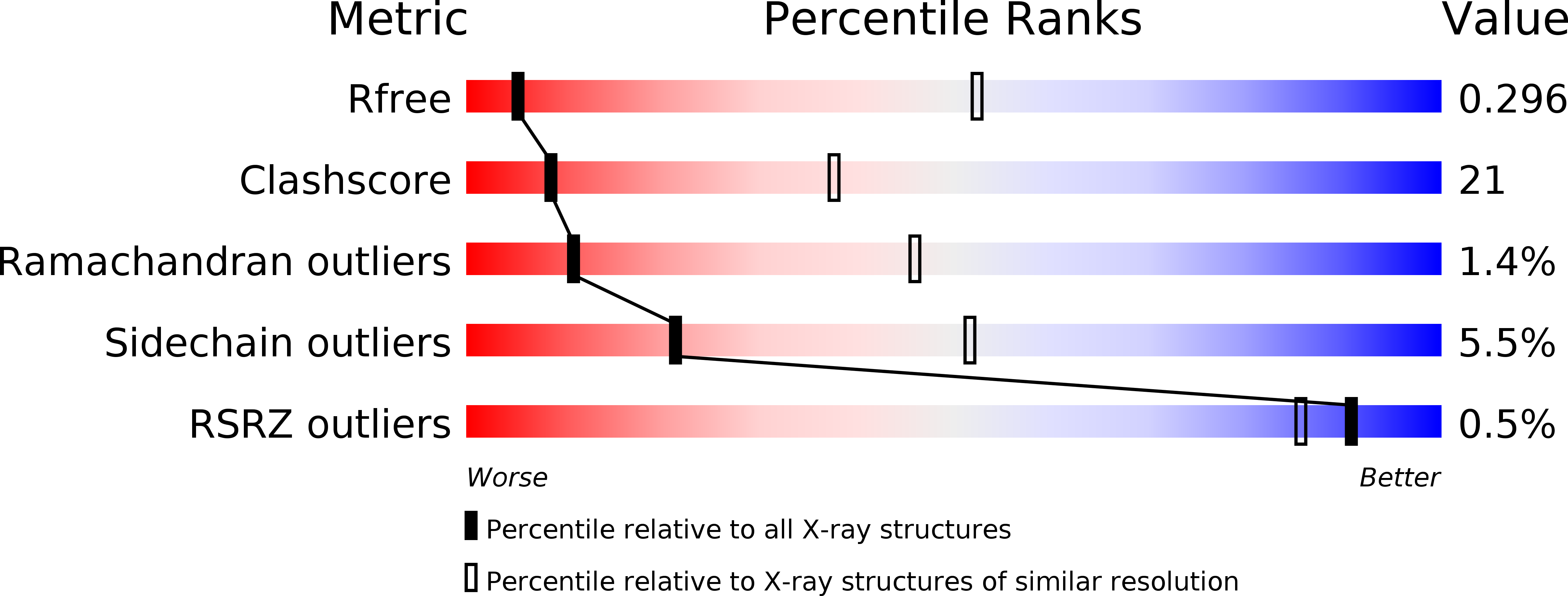
3T1P image
Deposition Date 2011-07-22
Release Date 2011-08-17
Last Version Date 2024-11-06
Entry Detail
PDB ID:
3T1P
Title:
Crystal structure of an alpha-1-antitrypsin trimer
Biological Source:
Source Organism:
Homo sapiens (Taxon ID: 9606)
Host Organism:
Method Details:
Experimental Method:
Resolution:
3.90 Å
R-Value Free:
0.29
R-Value Work:
0.23
R-Value Observed:
0.23
Space Group:
P 41 3 2
Macromolecular Entities
Structures with similar UniProt ID
Protein Blast
Polymer Type:polypeptide(L)
Molecule:Alpha-1-antitrypsin
Gene (Uniprot):SERPINA1
Mutagens:C256S, S316C, T363C, M382R
Chain IDs:A
Chain Length:371
Number of Molecules:1
Biological Source:Homo sapiens
Ligand Molecules
Primary Citation
Molecular basis of alpha 1-antitrypsin deficiency revealed by the structure of a domain-swapped trimer.
EMBO Rep. 12 1011 1017 (2011)
PMID: 21909074 DOI: 10.1038/embor.2011.171

Abstact

α(1)-Antitrypsin (α1AT) deficiency is a disease with multiple manifestations, including cirrhosis and emphysema, caused by the accumulation of stable polymers of mutant protein in the endoplasmic reticulum of hepatocytes. However, the molecular basis of misfolding and polymerization remain unknown. We produced and crystallized a trimeric form of α1AT that is recognized by an antibody specific for the pathological polymer. Unexpectedly, this structure reveals a polymeric linkage mediated by domain swapping the carboxy-terminal 34 residues. Disulphide-trapping and antibody-binding studies further demonstrate that runaway C-terminal domain swapping, rather than the s4A/s5A domain swap previously proposed, underlies polymerization of the common Z-mutant of α1AT in vivo.

Legend

Protein

Chemical

Disease

Primary Citation of related structures