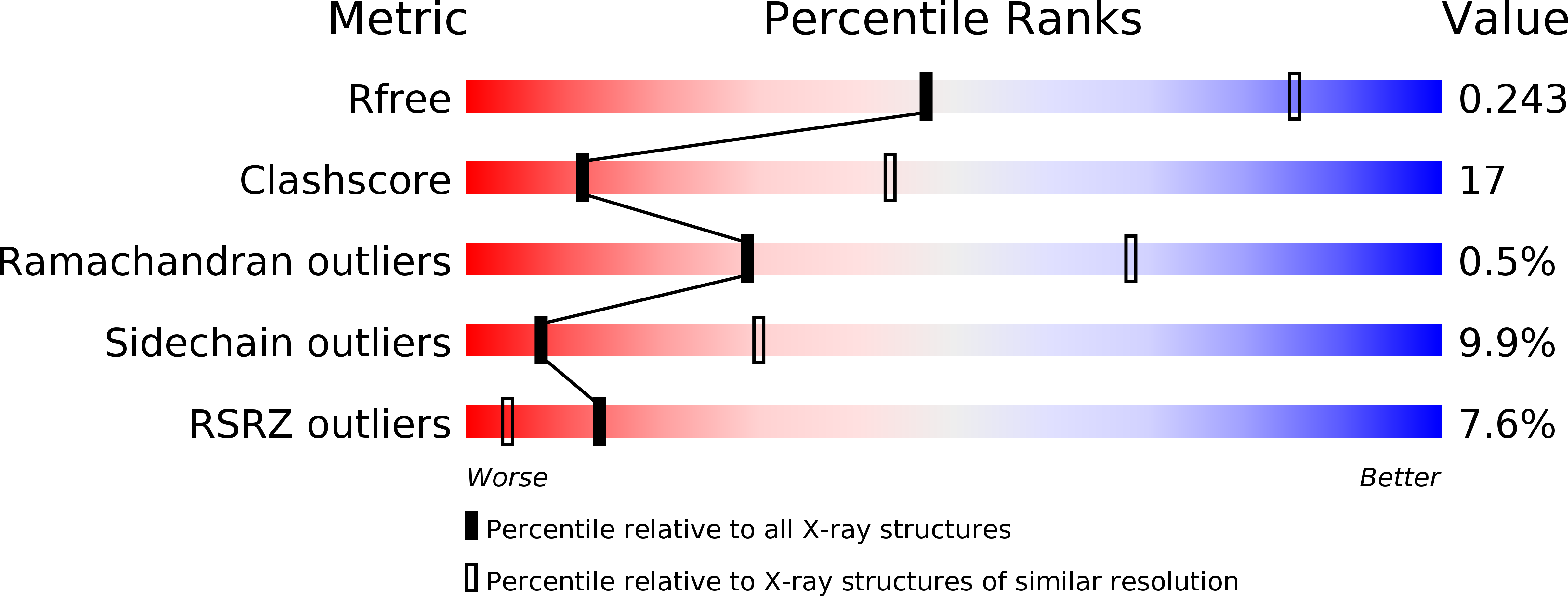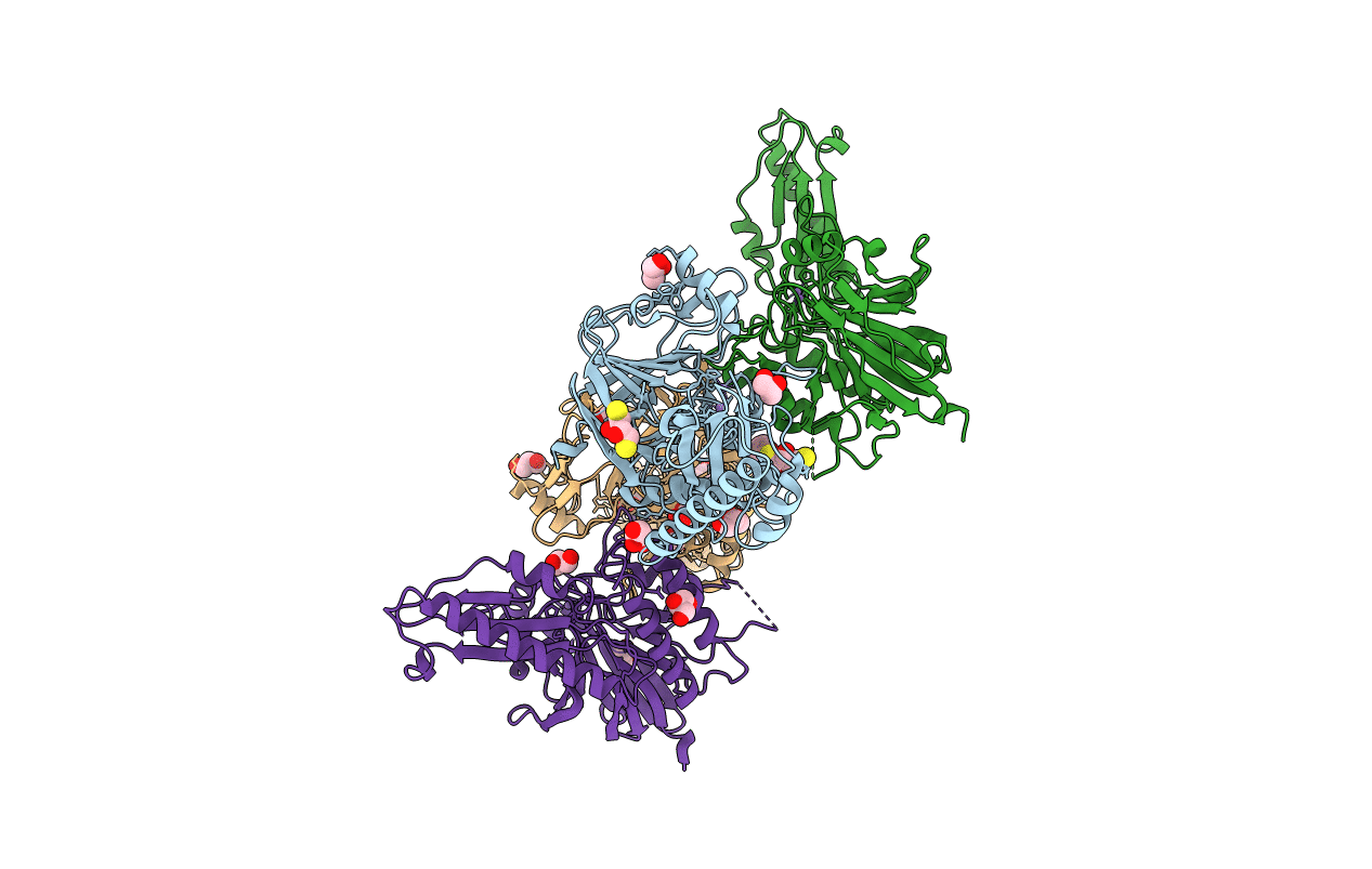
3T1I image
Deposition Date 2011-07-22
Release Date 2011-11-30
Last Version Date 2024-11-20
Entry Detail
PDB ID:
3T1I
Keywords:
Title:
Crystal Structure of Human Mre11: Understanding Tumorigenic Mutations
Biological Source:
Source Organism:
Homo sapiens (Taxon ID: 9606)
Host Organism:
Method Details:
Experimental Method:
Resolution:
3.00 Å
R-Value Free:
0.25
R-Value Work:
0.22
R-Value Observed:
0.22
Space Group:
P 21 21 21
Macromolecular Entities
Structures with similar UniProt ID
Protein Blast
Polymer Type:polypeptide(L)
Molecule:Double-strand break repair protein MRE11A
Gene (Uniprot):MRE11
Chain IDs:A, B, C, D
Chain Length:431
Number of Molecules:4
Biological Source:Homo sapiens
Modified Residue
Compound ID Chain ID Parent Comp ID Details 2D Image
MSE A MET SELENOMETHIONINE
Primary Citation
Crystal structure of human mre11: understanding tumorigenic mutations
Structure 19 1591 1602 (2011)
PMID: 22078559 DOI: 10.1016/j.str.2011.09.010

Abstact

Mre11 plays an important role in repairing damaged DNA by cleaving broken ends and by providing a platform for other DNA repair proteins. Various Mre11 mutations have been identified in several types of cancer. We have determined the crystal structure of the human Mre11 core (hMre11), which contains the nuclease and capping domains. hMre11 dimerizes through the interfaces between loop β3-α3 from one Mre11 and loop β4-β5 from another Mre11, and between loop α2-β3 from one Mre11 and helices α2 and α3 from another Mre11, and assembles into a completely different dimeric architecture compared with bacterial or archaeal Mre11 homologs. Nbs1 binds to the region containing loop α2-β3 which participates in dimerization. The hMre11 structure in conjunction with biochemical analyses reveals that many tumorigenic mutations are primarily associated with Nbs1 binding and partly with nuclease activities, providing a framework for understanding how mutations inactivate Mre11.

Legend

Protein

Chemical

Disease

Primary Citation of related structures