3T15 image
Deposition Date 2011-07-21
Release Date 2011-11-09
Last Version Date 2024-02-28
Entry Detail
PDB ID:
3T15
Keywords:
Title:
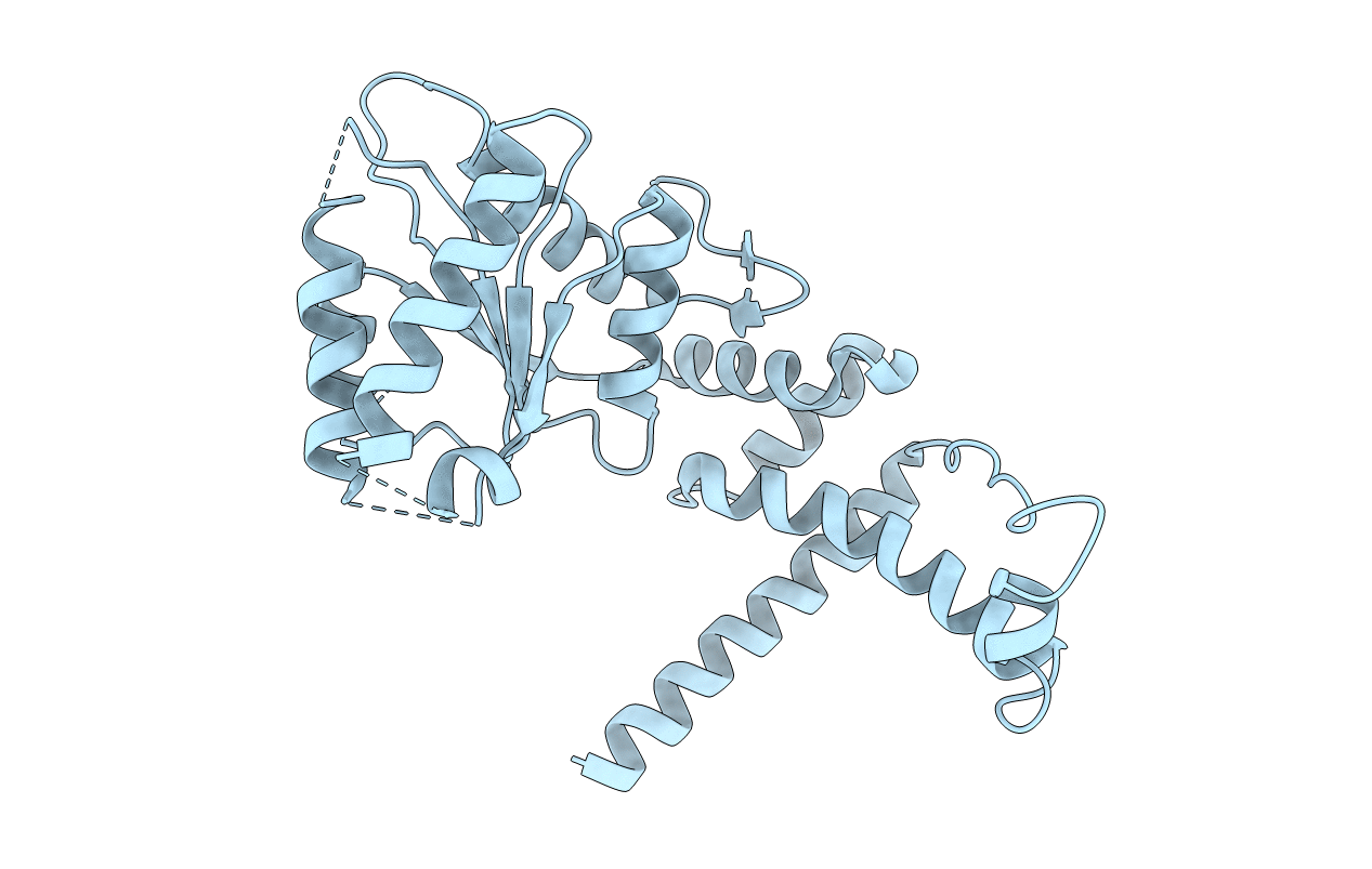
Structure of green-type Rubisco activase from tobacco
Biological Source:
Source Organism:
Host Organism:
Method Details:
Experimental Method:
Resolution:
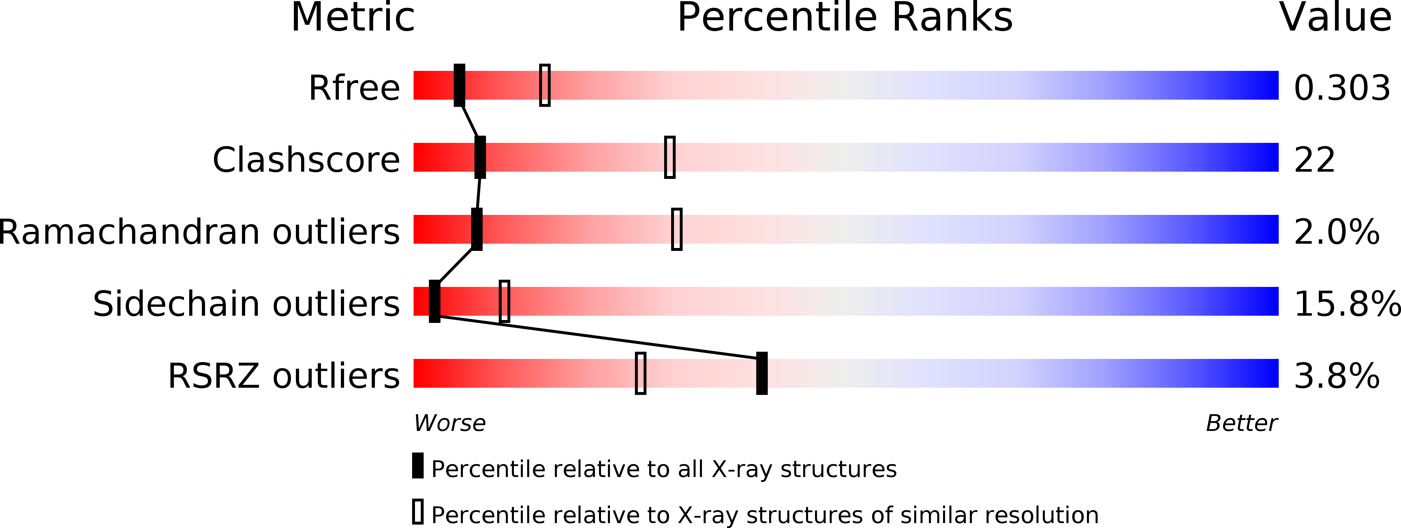
2.95 Å
R-Value Free:
0.29
R-Value Work:
0.22
R-Value Observed:
0.22
Space Group:
P 65
Macromolecular Entities
Structures with similar UniProt ID
Protein Blast
Polymer Type:polypeptide(L)
Molecule:Ribulose bisphosphate carboxylase/oxygenase activase 1, chloroplastic
Chain IDs:A
Chain Length:293
Number of Molecules:1
Biological Source:Nicotiana tabacum
Ligand Molecules
Primary Citation
Structure of green-type Rubisco activase from tobacco.
Nat.Struct.Mol.Biol. 18 1366 1370 (2011)
PMID: 22056769 DOI: 10.1038/nsmb.2171

Abstact

Rubisco, the enzyme that catalyzes the fixation of atmospheric CO(2) in photosynthesis, is subject to inactivation by inhibitory sugar phosphates. Here we report the 2.95-Å crystal structure of Nicotiana tabacum Rubisco activase (Rca), the enzyme that facilitates the removal of these inhibitors. Rca from tobacco has a classical AAA(+)-protein domain architecture. Although Rca populates a range of oligomeric states when in solution, it forms a helical arrangement with six subunits per turn when in the crystal. However, negative-stain electron microscopy of the active mutant R294V suggests that Rca functions as a hexamer. The residues determining species specificity for Rubisco are located in a helical insertion of the C-terminal domain and probably function in conjunction with the N-domain in Rubisco recognition. Loop segments exposed toward the central pore of the hexamer are required for the ATP-dependent remodeling of Rubisco, resulting in the release of inhibitory sugar.

Legend

Protein

Chemical

Disease

Primary Citation of related structures