3T0Y image
Deposition Date 2011-07-20
Release Date 2012-05-02
Last Version Date 2024-11-20
Entry Detail
PDB ID:
3T0Y
Title:
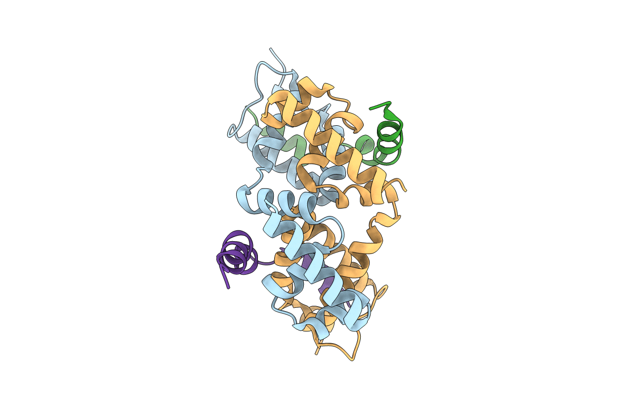
Structure of the PhyR anti-anti-sigma domain bound to the anti-sigma factor, NepR
Biological Source:
Source Organism:
Host Organism:
Method Details:
Experimental Method:
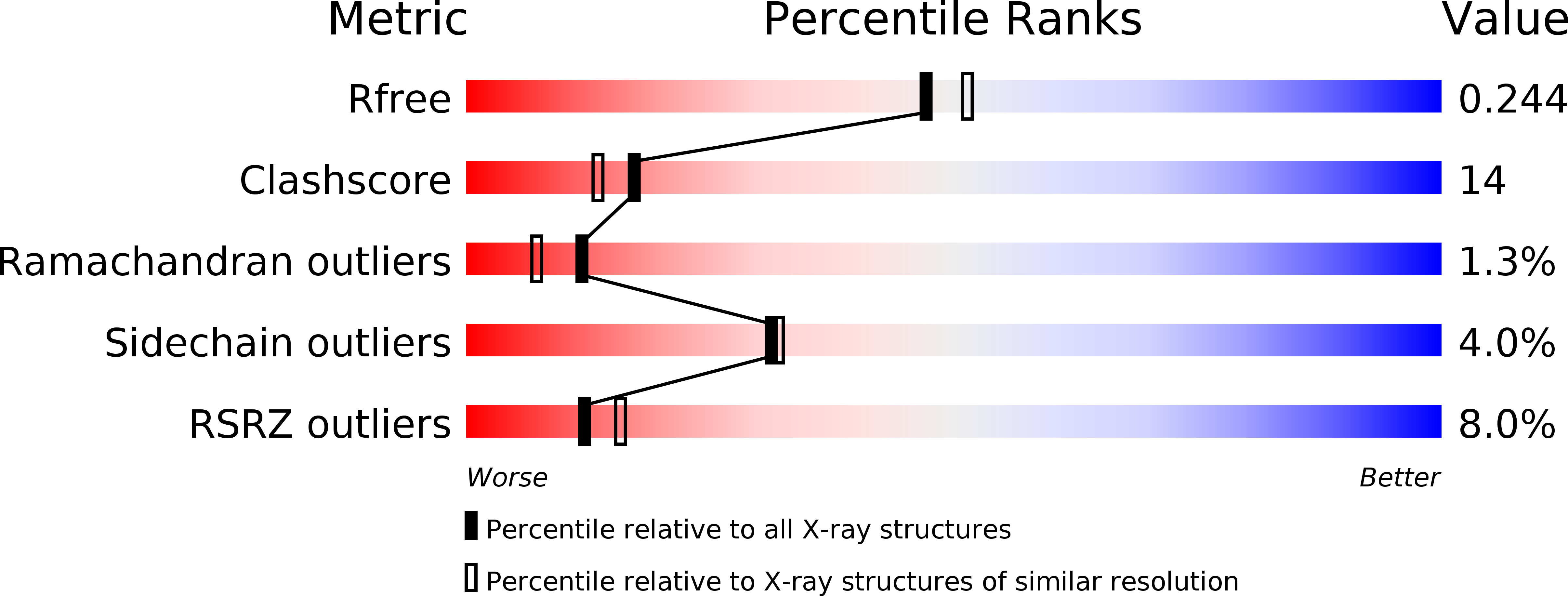
Resolution:
2.10 Å
R-Value Free:
0.25
R-Value Work:
0.20
R-Value Observed:
0.20
Space Group:
C 2 2 21
Macromolecular Entities
Structures with similar UniProt ID
Protein Blast
Polymer Type:polypeptide(L)
Molecule:Response regulator
Chain IDs:A, C
Chain Length:142
Number of Molecules:2
Biological Source:Caulobacter vibrioides
Structures with similar UniProt ID
Protein Blast
Polymer Type:polypeptide(L)
Molecule:NepR
Chain IDs:B, D
Chain Length:68
Number of Molecules:2
Biological Source:Caulobacter vibrioides
Modified Residue
Compound ID Chain ID Parent Comp ID Details 2D Image
MSE A MET SELENOMETHIONINE
Primary Citation
Structural basis of a protein partner switch that regulates the general stress response of alpha-proteobacteria
Proc.Natl.Acad.Sci.USA 109 E1415 E1423 (2012)
PMID: 22550172 DOI: 10.1073/pnas.1116887109

Abstact

α-Proteobacteria uniquely integrate features of two-component signal transduction (TCS) and alternative sigma factor (σ) regulation to control transcription in response to general stress. The core of this regulatory system is the PhyR protein, which contains a σ-like (SL) domain and a TCS receiver domain. Aspartyl phosphorylation of the PhyR receiver in response to stress signals promotes binding of the anti-σ factor, NepR, to PhyR-SL. This mechanism, whereby NepR switches binding between its cognate σ factor and phospho-PhyR (PhyR∼P), controls transcription of the general stress regulon. We have defined the structural basis of the PhyR∼P/NepR interaction in Caulobacter crescentus and characterized the effect of aspartyl phosphorylation on PhyR structure by molecular dynamics simulations. Our data support a model in which phosphorylation of the PhyR receiver domain promotes its dissociation from the PhyR-SL domain, which exposes the NepR binding site. A highly dynamic loop-helix region (α3-α4) of the PhyR-SL domain plays an important role in PhyR∼P binding to NepR in vitro, and in stress-dependent activation of transcription in vivo. This study provides a foundation for understanding the protein-protein interactions and protein structural dynamics that underpin general stress adaptation in a large and metabolically diverse clade of the bacterial kingdom.

Legend

Protein

Chemical

Disease

Primary Citation of related structures
Feedback Form
Name
Email
Institute
Feedback