Deposition Date
2011-07-19
Release Date
2011-08-03
Last Version Date
2024-02-28
Entry Detail
PDB ID:
3SZY
Keywords:
Title:
Crystal Structure of Phosphonoacetate hydrolase from Sinorhizobium meliloti 1021 in APO form
Biological Source:
Source Organism(s):
Sinorhizobium meliloti (Taxon ID: 266834)
Expression System(s):
Method Details:
Experimental Method:
Resolution:
1.35 Å
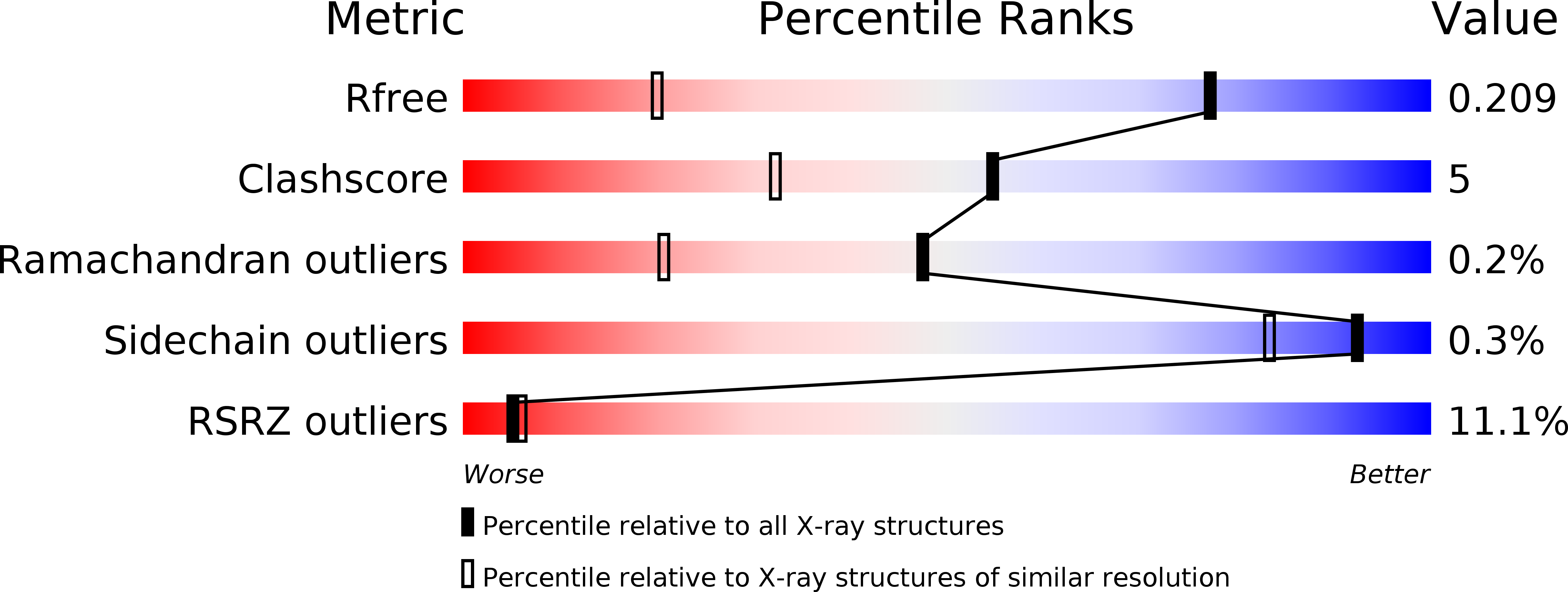
R-Value Free:
0.20
R-Value Work:
0.19
R-Value Observed:
0.19
Space Group:
P 43 21 2