Deposition Date
2011-07-18
Release Date
2012-02-15
Last Version Date
2023-11-01
Entry Detail
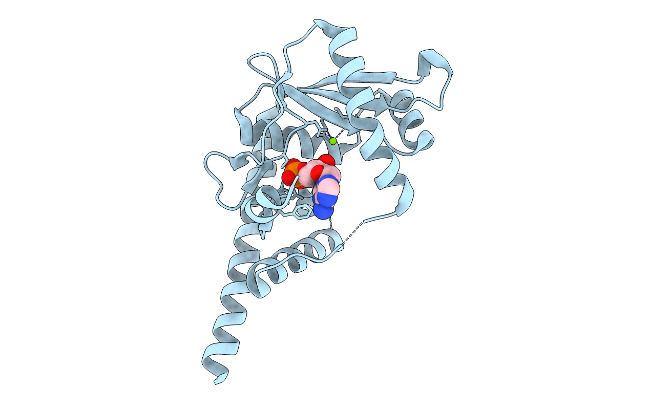
Biological Source:
Source Organism(s):
Laribacter hongkongensis (Taxon ID: 557598)
Expression System(s):
Method Details:
Experimental Method:
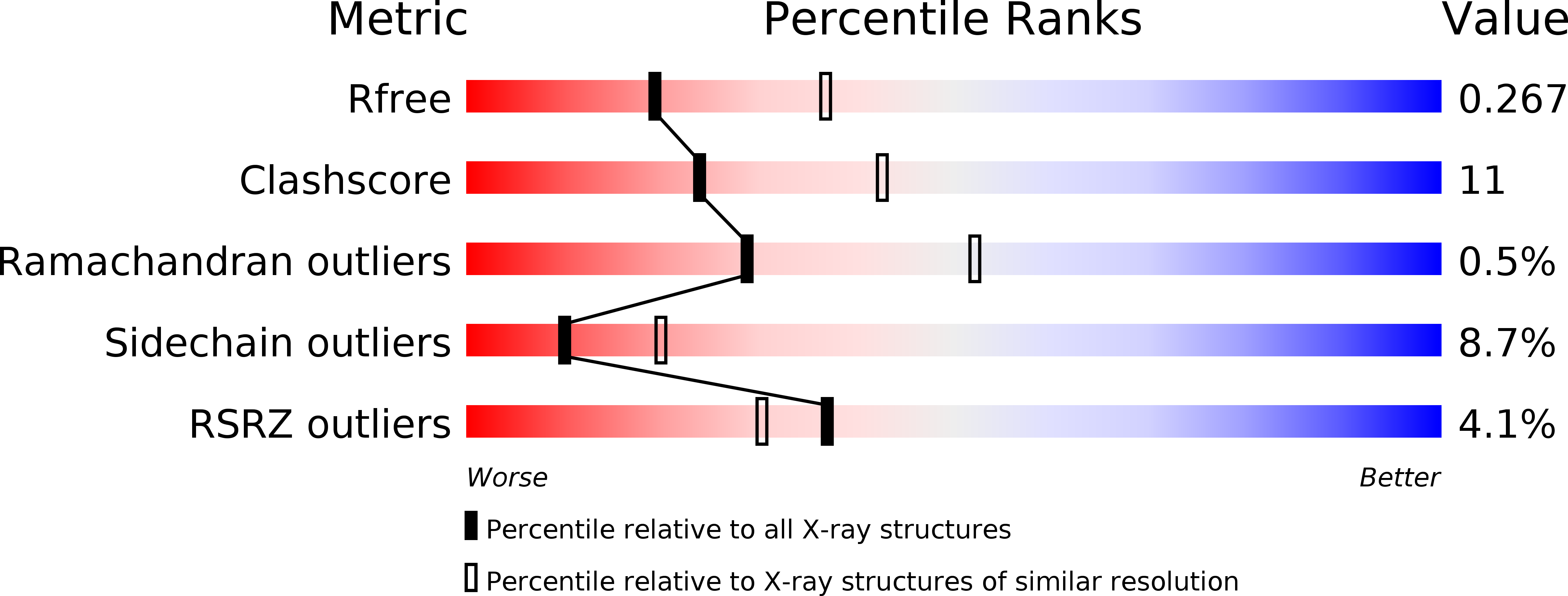
Resolution:
2.59 Å
R-Value Free:
0.26
R-Value Work:
0.20
R-Value Observed:
0.21
Space Group:
P 63