3SYW image
Deposition Date 2011-07-18
Release Date 2012-04-25
Last Version Date 2024-03-13
Entry Detail
PDB ID:
3SYW
Keywords:
Title:
Crystal Structure of the Triplet Repeat in Myotonic Dystrophy Reveals Heterogeneous 1x1 Nucleotide UU Internal Loop Conformations
Biological Source:
Source Organism:
(Taxon ID: )
Method Details:
Experimental Method:
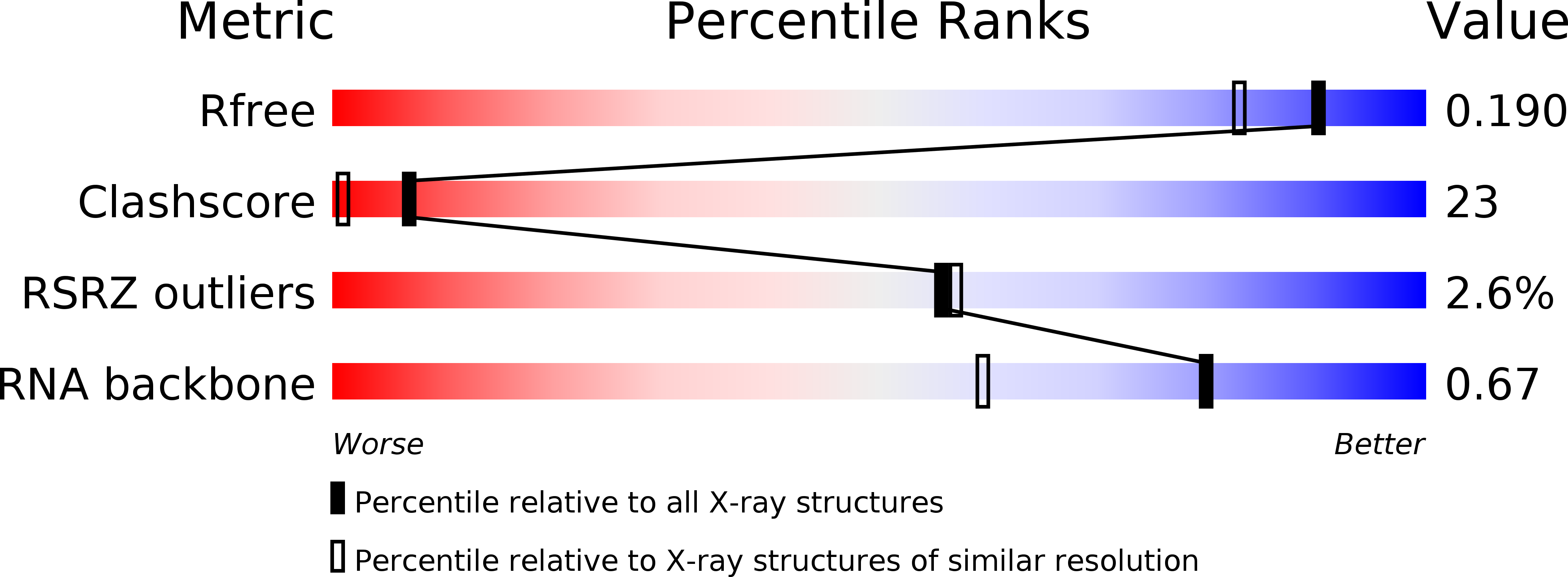
Resolution:
1.57 Å
R-Value Free:
0.18
R-Value Work:
0.16
R-Value Observed:
0.16
Space Group:
H 3
Macromolecular Entities
Polymer Type:polyribonucleotide
Molecule:RNA (5'-R(*UP*UP*GP*GP*GP*CP*CP*UP*GP*CP*UP*GP*CP*UP*GP*GP*UP*CP*C)-3')
Chain IDs:A, B
Chain Length:19
Number of Molecules:2
Biological Source:
Ligand Molecules
Primary Citation
Crystal Structure of the Triplet Repeat in Myotonic Dystrophy Reveals Heterogeneous 1x1 Nucleotide UU Internal Loop Conformations
Biochemistry 50 9928 9935 (2011)
PMID: 21988728 DOI: 10.1021/bi2013068

Abstact

RNA internal loops often display a variety of conformations in solution. Herein, we visualize conformational heterogeneity in the context of the 5'CUG/3'GUC repeat motif present in the RNA that causes myotonic dystrophy type 1 (DM1). Specifically, two crystal structures of a model DM1 triplet repeating construct, 5'r[UUGGGC(CUG)(3)GUCC](2), refined to 2.20 and 1.52 Å resolution are disclosed. Here, differences in the orientation of the 5' dangling UU end between the two structures induce changes in the backbone groove width, which reveals that noncanonical 1 × 1 nucleotide UU internal loops can display an ensemble of pairing conformations. In the 2.20 Å structure, CUGa, the 5' UU forms a one hydrogen-bonded pair with a 5' UU of a neighboring helix in the unit cell to form a pseudoinfinite helix. The central 1 × 1 nucleotide UU internal loop has no hydrogen bonds, while the terminal 1 × 1 nucleotide UU internal loops each form a one-hydrogen bond pair. In the 1.52 Å structure, CUGb, the 5' UU dangling end is tucked into the major groove of the duplex. While the canonically paired bases show no change in base pairing, in CUGb the terminal 1 × 1 nucleotide UU internal loops now form two hydrogen-bonded pairs. Thus, the shift in the major groove induced by the 5' UU dangling end alters noncanonical base patterns. Collectively, these structures indicate that 1 × 1 nucleotide UU internal loops in DM1 may sample multiple conformations in vivo. This observation has implications for the recognition of this RNA, and other repeating transcripts, by protein and small molecule ligands.

Legend

Protein

Chemical

Disease

Primary Citation of related structures