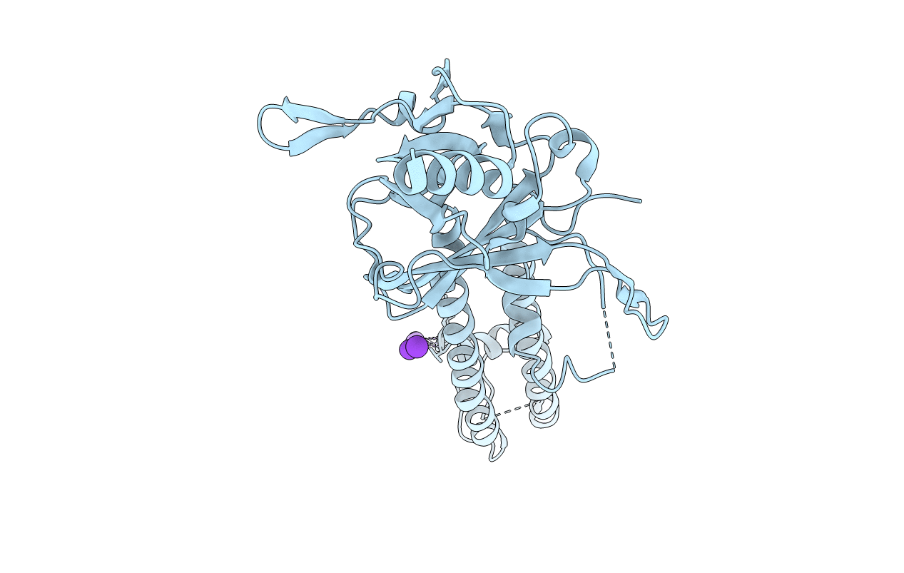
3SYC image
Deposition Date 2011-07-16
Release Date 2011-10-12
Last Version Date 2024-10-30
Entry Detail
PDB ID:
3SYC
Keywords:
Title:
Crystal structure of the G protein-gated inward rectifier K+ channel GIRK2 (Kir3.2) D228N mutant
Biological Source:
Source Organism:
Mus musculus (Taxon ID: 10090)
Host Organism:
Method Details:
Experimental Method:
Resolution:
3.41 Å
R-Value Free:
0.27
R-Value Work:
0.25
R-Value Observed:
0.25
Space Group:
P 4 21 2
Macromolecular Entities
Polymer Type:polypeptide(L)
Molecule:G protein-activated inward rectifier potassium channel 2
Gene (Uniprot):Kcnj6
Mutations:D228N
Chain IDs:A
Chain Length:340
Number of Molecules:1
Biological Source:Mus musculus
Primary Citation
Crystal Structure of the Mammalian GIRK2 K(+) Channel and Gating Regulation by G Proteins, PIP(2), and Sodium.
Cell(Cambridge,Mass.) 147 199 208 (2011)
PMID: 21962516 DOI: 10.1016/j.cell.2011.07.046

Abstact

G protein-gated K(+) channels (Kir3.1-Kir3.4) control electrical excitability in many different cells. Among their functions relevant to human physiology and disease, they regulate the heart rate and govern a wide range of neuronal activities. Here, we present the first crystal structures of a G protein-gated K(+) channel. By comparing the wild-type structure to that of a constitutively active mutant, we identify a global conformational change through which G proteins could open a G loop gate in the cytoplasmic domain. The structures of both channels in the absence and presence of PIP(2) suggest that G proteins open only the G loop gate in the absence of PIP(2), but in the presence of PIP(2) the G loop gate and a second inner helix gate become coupled, so that both gates open. We also identify a strategically located Na(+) ion-binding site, which would allow intracellular Na(+) to modulate GIRK channel activity. These data provide a structural basis for understanding multiligand regulation of GIRK channel gating.

Legend

Protein

Chemical

Disease

Primary Citation of related structures