Deposition Date
2011-07-15
Release Date
2012-02-08
Last Version Date
2023-09-13
Entry Detail
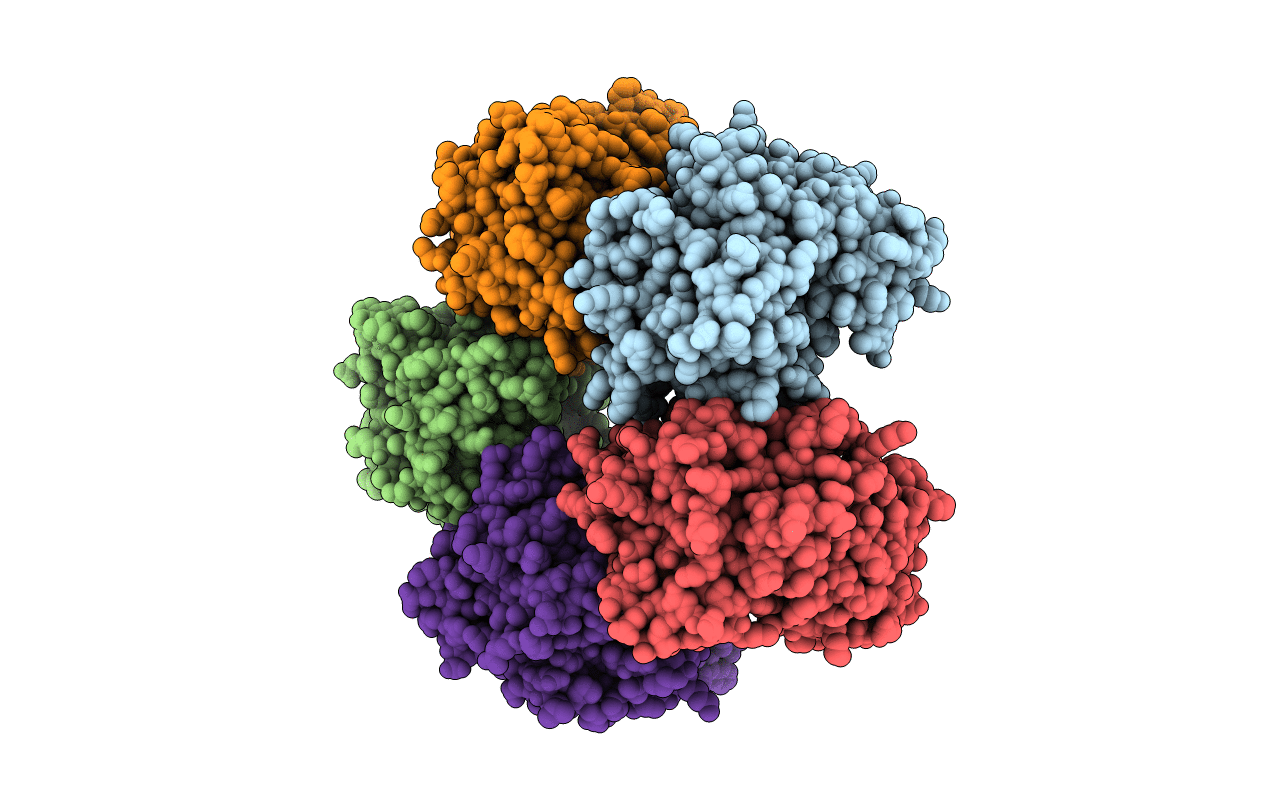
PDB ID:
3SXP
Keywords:
Title:
Crystal Structure of Helicobacter pylori ADP-L-glycero-D-manno-heptose-6-epimerase (rfaD, HP0859)
Biological Source:
Source Organism(s):
Helicobacter pylori (Taxon ID: 563041)
Expression System(s):
Method Details:
Experimental Method:
Resolution:
2.55 Å
R-Value Free:
0.27
R-Value Work:
0.23
R-Value Observed:
0.23
Space Group:
I 1 2 1