3SWS image
Deposition Date 2011-07-14
Release Date 2012-07-25
Last Version Date 2023-09-13
Entry Detail
PDB ID:
3SWS
Title:
Crystal Structure of the Quinone Form of Methylamine Dehydrogenase in Complex with the Diferric Form of MauG
Biological Source:
Source Organism:
Method Details:
Experimental Method:
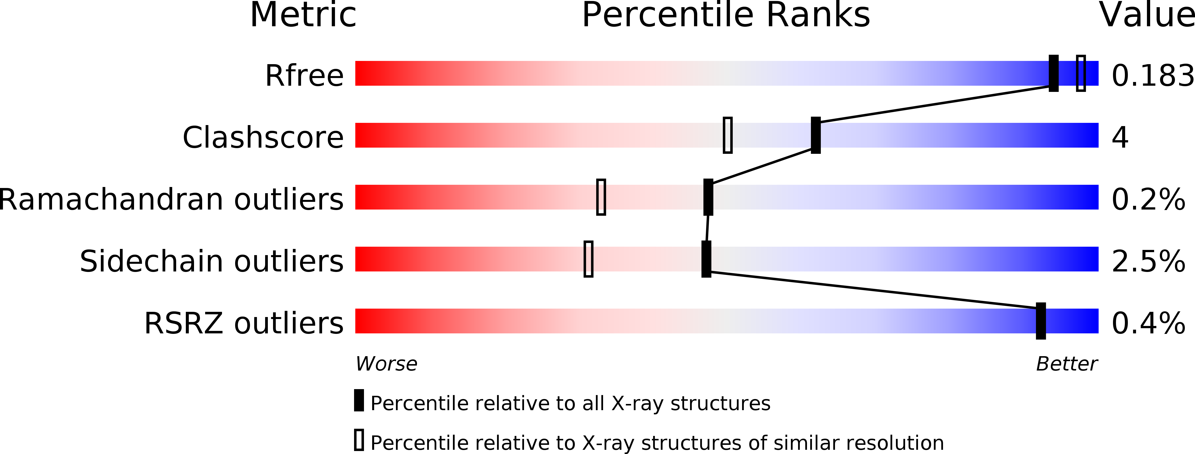
Resolution:
1.86 Å
R-Value Free:
0.17
R-Value Work:
0.13
R-Value Observed:
0.13
Space Group:
P 1
Macromolecular Entities
Structures with similar UniProt ID
Protein Blast
Polymer Type:polypeptide(L)
Molecule:Methylamine utilization protein MauG
Gene (Uniprot):mauG
Chain IDs:A, B
Chain Length:373
Number of Molecules:2
Biological Source:Paracoccus denitrificans
Structures with similar UniProt ID
Protein Blast
Polymer Type:polypeptide(L)
Molecule:Methylamine dehydrogenase light chain
Chain IDs:C, E
Chain Length:137
Number of Molecules:2
Biological Source:Paracoccus denitrificans
Structures with similar UniProt ID
Protein Blast
Polymer Type:polypeptide(L)
Molecule:Methylamine dehydrogenase heavy chain
Gene (Uniprot):Pden_4730
Chain IDs:D, F
Chain Length:386
Number of Molecules:2
Biological Source:Paracoccus denitrificans
Modified Residue
Compound ID Chain ID Parent Comp ID Details 2D Image
TRQ C TRP ?
Primary Citation
Crystal Structure of the Quinone Form of Methylamine Dehydrogenase in Complex with the Diferric Form of MauG
To be Published ? ? ? (?)
Primary Citation of related structures
Feedback Form
Name
Email
Institute
Feedback