3SVI image
Deposition Date 2011-07-12
Release Date 2011-08-10
Last Version Date 2024-11-20
Entry Detail
PDB ID:
3SVI
Title:
Structure of the Pto-binding domain of HopPmaL generated by limited thermolysin digestion
Biological Source:
Source Organism:
Host Organism:
Method Details:
Experimental Method:
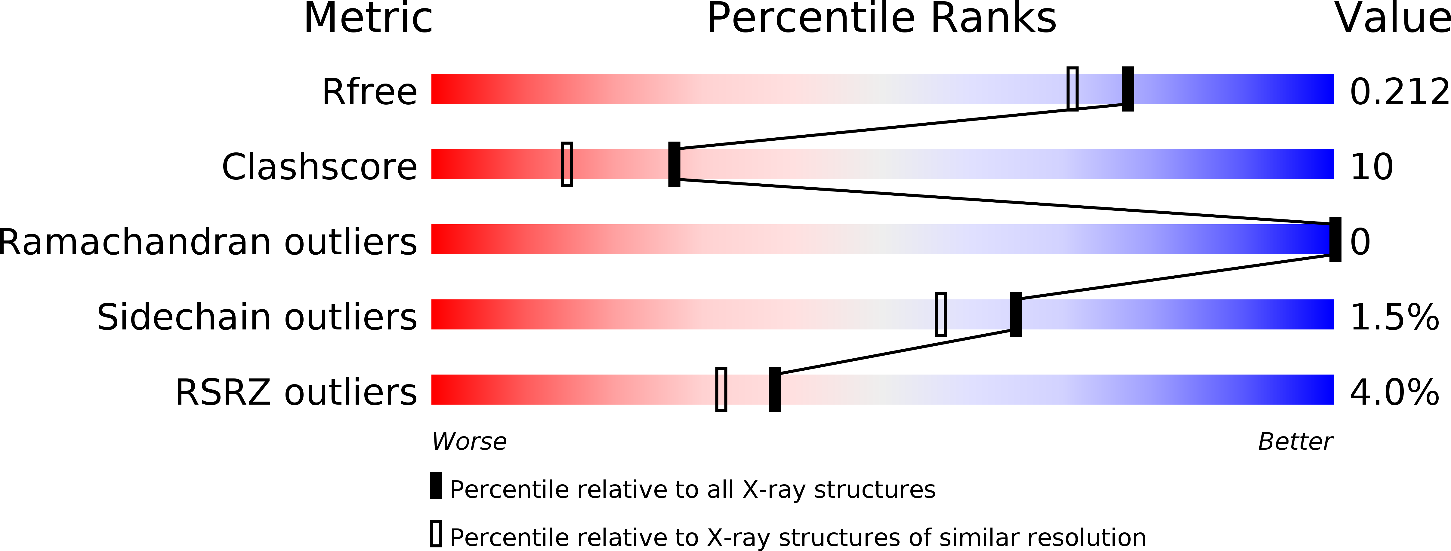
Resolution:
1.80 Å
R-Value Free:
0.21
R-Value Work:
0.18
R-Value Observed:
0.18
Space Group:
P 41 21 2
Macromolecular Entities
Polymer Type:polypeptide(L)
Molecule:Type III effector HopAB2
Chain IDs:A
Chain Length:83
Number of Molecules:1
Biological Source:Pseudomonas syringae
Modified Residue
Compound ID Chain ID Parent Comp ID Details 2D Image
MSE A MET SELENOMETHIONINE
Primary Citation
Structural Analysis of HopPmaL Reveals the Presence of a Second Adaptor Domain Common to the HopAB Family of Pseudomonas syringae Type III Effectors.
Biochemistry 51 1 3 (2012)
PMID: 22191472 DOI: 10.1021/bi2013883

Abstact

HopPmaL is a member of the HopAB family of type III effectors present in the phytopathogen Pseudomonas syringae. Using both X-ray crystallography and solution nuclear magnetic resonance, we demonstrate that HopPmaL contains two structurally homologous yet functionally distinct domains. The N-terminal domain corresponds to the previously described Pto-binding domain, while the previously uncharacterised C-terminal domain spans residues 308-385. While structurally similar, these domains do not share significant sequence similarity and most importantly demonstrate significant differences in key residues involved in host protein recognition, suggesting that each of them targets a different host protein.

Legend

Protein

Chemical

Disease

Primary Citation of related structures