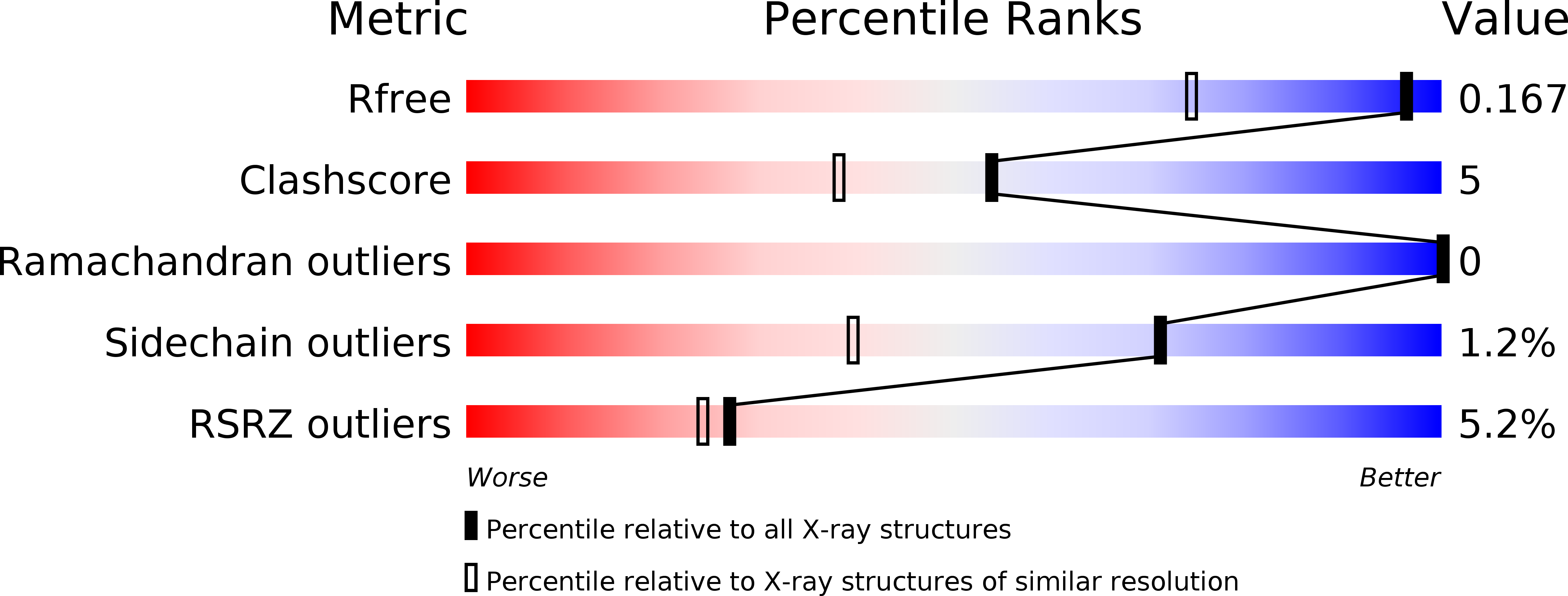
3SV2 image
Deposition Date 2011-07-12
Release Date 2012-06-20
Last Version Date 2024-11-20
Entry Detail
PDB ID:
3SV2
Title:
Human Thrombin In Complex With UBTHR105
Biological Source:
Source Organism:
Hirudo medicinalis (Taxon ID: 6421)
Homo sapiens (Taxon ID: 9606)
Method Details:
Experimental Method:
Resolution:
1.30 Å
R-Value Free:
0.16
R-Value Work:
0.14
R-Value Observed:
0.14
Space Group:
C 1 2 1
Macromolecular Entities
Polymer Type:polypeptide(L)
Molecule:Thrombin heavy chain
Gene (Uniprot):F2
Chain IDs:B (auth: H)
Chain Length:259
Number of Molecules:1
Biological Source:Homo sapiens
Polymer Type:polypeptide(L)
Molecule:Hirudin variant-2
Chain IDs:C (auth: I)
Chain Length:13
Number of Molecules:1
Biological Source:Hirudo medicinalis
Polymer Type:polypeptide(L)
Molecule:Thrombin light chain
Gene (Uniprot):F2
Chain IDs:A (auth: L)
Chain Length:36
Number of Molecules:1
Biological Source:Homo sapiens
Modified Residue
Compound ID Chain ID Parent Comp ID Details 2D Image
ASN B ASN GLYCOSYLATION SITE
TYS C TYR O-SULFO-L-TYROSINE
Peptide-like Molecules
PRD_000795
Primary Citation
Impact of ligand and protein desolvation on ligand binding to the S1 pocket of thrombin
J.Mol.Biol. 418 350 366 (2012)
PMID: 22366545 DOI: 10.1016/j.jmb.2012.01.054

Abstact

In the present study, we investigate the impact of a tightly bound water molecule on ligand binding in the S1 pocket of thrombin. The S1 pocket contains a deeply buried deprotonated aspartate residue (Asp189) that is, due to its charged state, well hydrated in the uncomplexed state. We systematically studied the importance of this water molecule by evaluating a series of ligands that contains pyridine-type P1 side chains that could potentially alter the binding properties of this water molecule. All of the pyridine derivatives retain the original hydration state albeit sometimes with a slight perturbance. In order to prevent a direct H-bond formation with Asp189, and to create a permanent positive charge on the P1 side chain that is positioned adjacent to the Asp189 carboxylate anion, we methylated the pyridine nitrogen. This methylation resulted in displacement of water but was accompanied by a loss in binding affinity. Quantum chemical calculations of the ligand solvation free energy showed that the positively charged methylpyridinium derivatives suffer a large penalty of desolvation upon binding. Consequently, they have a substantially less favorable enthalpy of binding. In addition to the ligand desolvation penalty, the hydration shell around Asp189 has to be overcome, which is achieved in nearly all pyridinium derivatives. Only for the ortho derivative is a partial population of a water next to Asp189 found. Possibly, the gain of electrostatic interactions between the charged P1 side chain and Asp189 helps to compensate for the desolvation penalty. In all uncharged pyridine derivatives, the solvation shell remains next to Asp189, partly mediating interactions between ligand and protein. In the case of the para-pyridine derivative, a strongly disordered cluster of water sites is observed between ligand and Asp189.

Legend

Protein

Chemical

Disease

Primary Citation of related structures