
Deposition Date
2011-07-11
Release Date
2012-06-06
Last Version Date
2024-11-20
Entry Detail
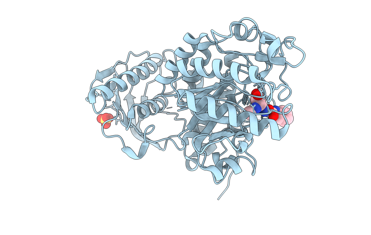
PDB ID:
3SUT
Keywords:
Title:
Crystal structure of beta-hexosaminidase from Paenibacillus sp. TS12 in complex with PUGNAc
Biological Source:
Source Organism(s):
Paenibacillus (Taxon ID: 192895)
Expression System(s):
Method Details:
Experimental Method:
Resolution:
1.90 Å
R-Value Free:
0.18
R-Value Work:
0.14
R-Value Observed:
0.14
Space Group:
C 2 2 21
