3STB image
Deposition Date 2011-07-09
Release Date 2011-11-02
Last Version Date 2024-11-20
Entry Detail
PDB ID:
3STB
Title:
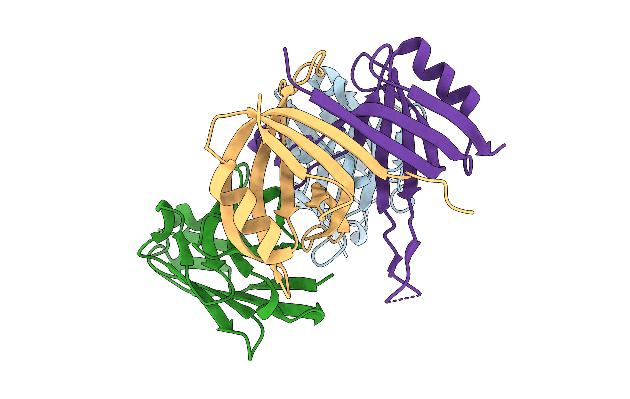
A complex of two editosome proteins and two nanobodies
Biological Source:
Source Organism:
Lama glama (Taxon ID: 9844)
Trypanosoma brucei (Taxon ID: 5691)
Host Organism:
Method Details:
Experimental Method:
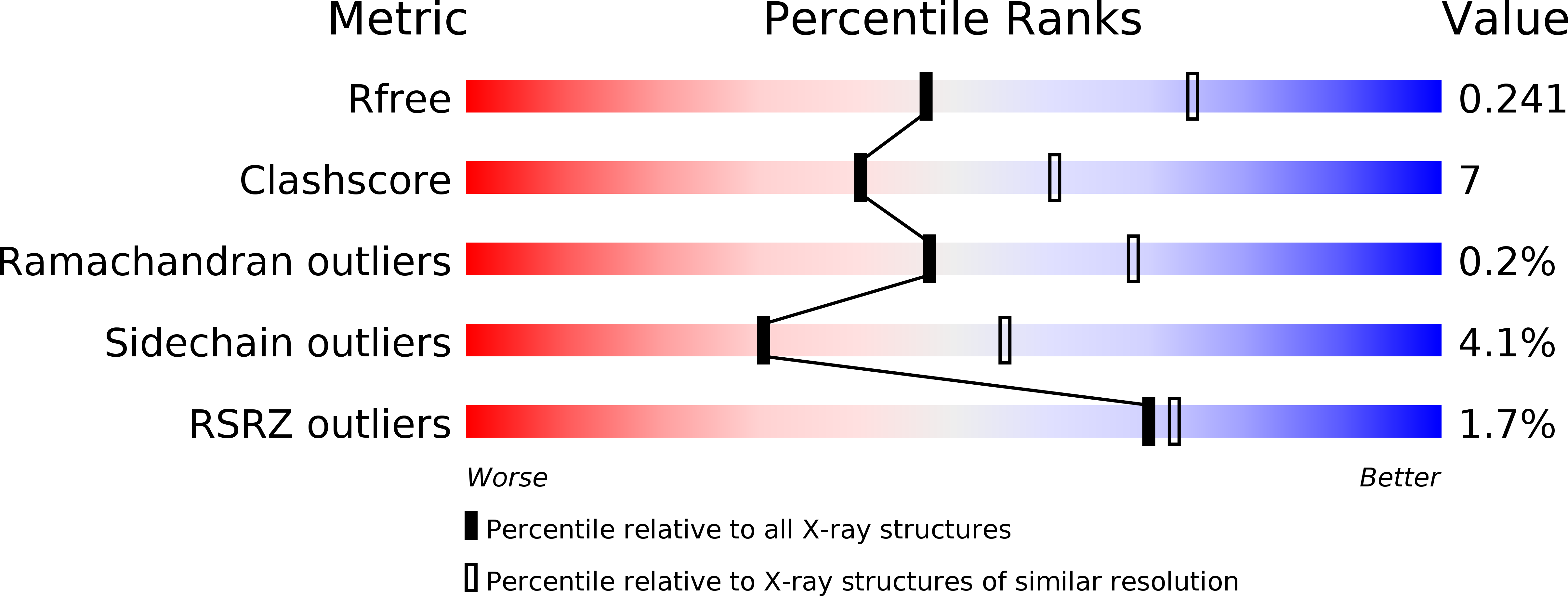
Resolution:
2.50 Å
R-Value Free:
0.23
R-Value Work:
0.19
R-Value Observed:
0.20
Space Group:
P 43 21 2
Macromolecular Entities
Polymer Type:polypeptide(L)
Molecule:single domain antibody VHH
Chain IDs:A, B
Chain Length:132
Number of Molecules:2
Biological Source:Lama glama
Structures with similar UniProt ID
Protein Blast
Polymer Type:polypeptide(L)
Molecule:RNA-editing complex protein MP42
Chain IDs:C
Chain Length:148
Number of Molecules:1
Biological Source:Trypanosoma brucei
Structures with similar UniProt ID
Protein Blast
Polymer Type:polypeptide(L)
Molecule:MP18 RNA editing complex protein
Gene (Uniprot):Tb10.70.2090
Chain IDs:D
Chain Length:145
Number of Molecules:1
Biological Source:Trypanosoma brucei
Primary Citation
Crystal structure of a heterodimer of editosome interaction proteins in complex with two copies of a cross-reacting nanobody.
Nucleic Acids Res. 40 1828 1840 (2012)
PMID: 22039098 DOI: 10.1093/nar/gkr867

Abstact

The parasite Trypanosoma brucei, the causative agent of sleeping sickness across sub-Saharan Africa, depends on a remarkable U-insertion/deletion RNA editing process in its mitochondrion. A approximately 20 S multi-protein complex, called the editosome, is an essential machinery for editing pre-mRNA molecules encoding the majority of mitochondrial proteins. Editosomes contain a common core of twelve proteins where six OB-fold interaction proteins, called A1-A6, play a crucial role. Here, we report the structure of two single-strand nucleic acid-binding OB-folds from interaction proteins A3 and A6 that surprisingly, form a heterodimer. Crystal growth required the assistance of an anti-A3 nanobody as a crystallization chaperone. Unexpectedly, this anti-A3 nanobody binds to both A3(OB) and A6, despite only ~40% amino acid sequence identity between the OB-folds of A3 and A6. The A3(OB)-A6 heterodimer buries 35% more surface area than the A6 homodimer. This is attributed mainly to the presence of a conserved Pro-rich loop in A3(OB). The implications of the A3(OB)-A6 heterodimer, and of a dimer of heterodimers observed in the crystals, for the architecture of the editosome are profound, resulting in a proposal of a 'five OB-fold center' in the core of the editosome.

Legend

Protein

Chemical

Disease

Primary Citation of related structures