3SOH image
Deposition Date 2011-06-30
Release Date 2011-07-27
Last Version Date 2023-09-13
Entry Detail
PDB ID:
3SOH
Keywords:
Title:
Architecture of the Flagellar Rotor
Biological Source:
Source Organism:
Host Organism:
Method Details:
Experimental Method:
Resolution:
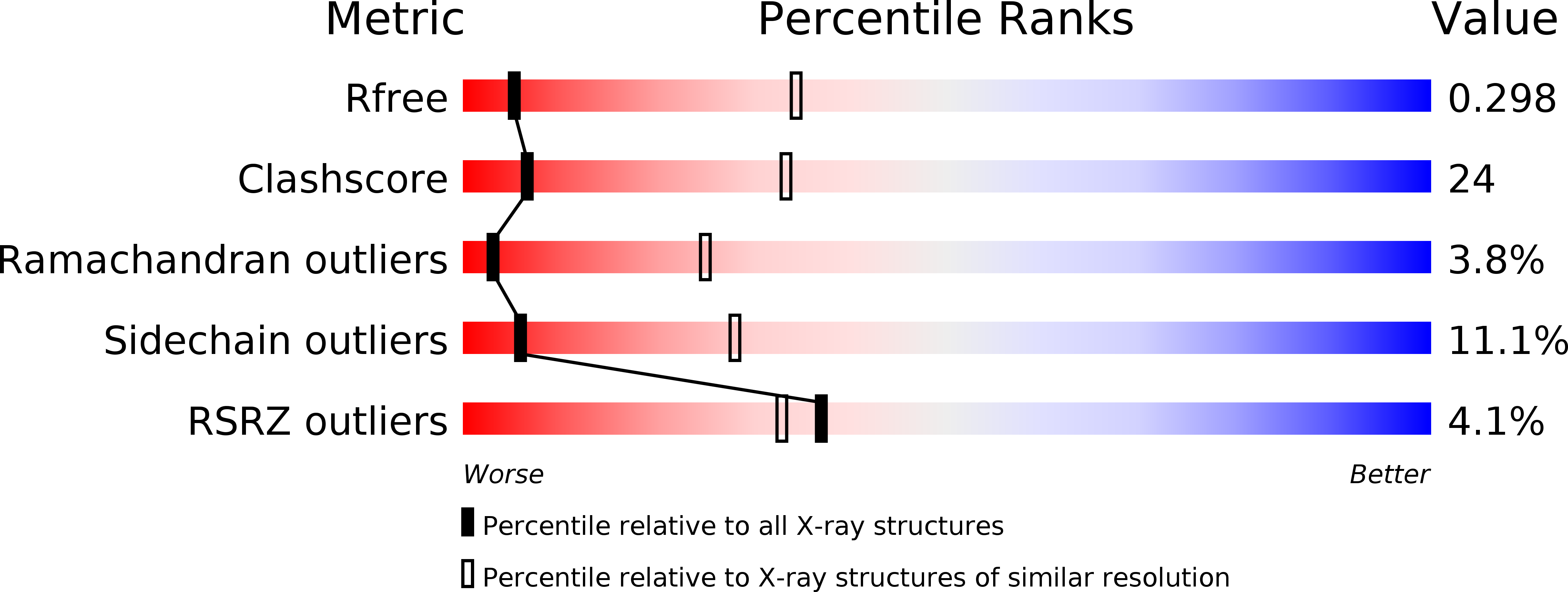
3.50 Å
R-Value Free:
0.30
R-Value Work:
0.25
R-Value Observed:
0.25
Space Group:
P 32 2 1
Macromolecular Entities
Polymer Type:polypeptide(L)
Molecule:Flagellar motor switch protein FliM
Gene (Uniprot):fliM
Chain IDs:A, C
Chain Length:188
Number of Molecules:2
Biological Source:Thermotoga maritima
Polymer Type:polypeptide(L)
Molecule:Flagellar motor switch protein FliG
Gene (Uniprot):fliG
Chain IDs:B, D
Chain Length:80
Number of Molecules:2
Biological Source:Thermotoga maritima
Primary Citation
Architecture of the flagellar rotor.
Embo J. 30 2962 2971 (2011)
PMID: 21673656 DOI: 10.1038/emboj.2011.188

Abstact

Rotation and switching of the bacterial flagellum depends on a large rotor-mounted protein assembly composed of the proteins FliG, FliM and FliN, with FliG most directly involved in rotation. The crystal structure of a complex between the central domains of FliG and FliM, in conjunction with several biochemical and molecular-genetic experiments, reveals the arrangement of the FliG and FliM proteins in the rotor. A stoichiometric mismatch between FliG (26 subunits) and FliM (34 subunits) is explained in terms of two distinct positions for FliM: one where it binds the FliG central domain and another where it binds the FliG C-terminal domain. This architecture provides a structural framework for addressing the mechanisms of motor rotation and direction switching and for unifying the large body of data on motor performance. Recently proposed alternative models of rotor assembly, based on a subunit contact observed in crystals, are not supported by experiment.

Legend

Protein

Chemical

Disease

Primary Citation of related structures