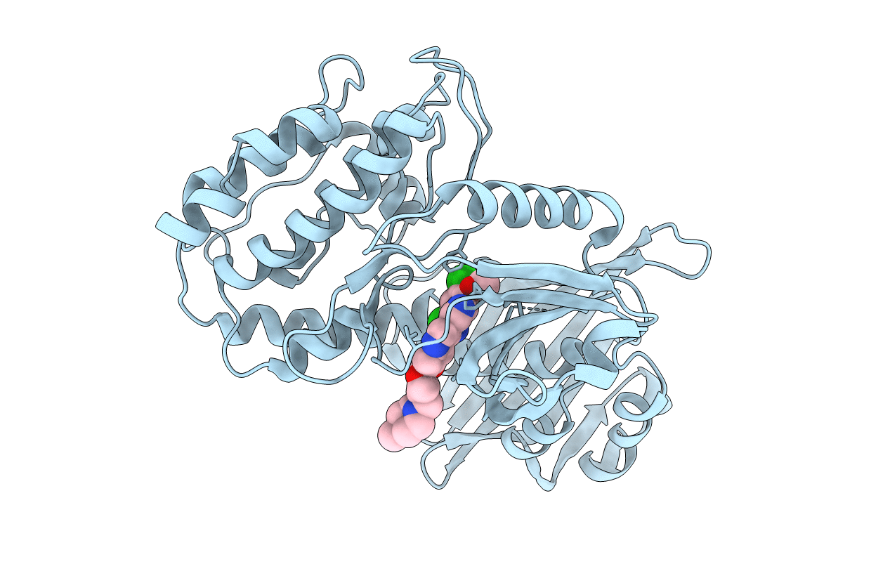
3SOA image
Deposition Date 2011-06-30
Release Date 2011-08-31
Last Version Date 2023-09-13
Entry Detail
PDB ID:
3SOA
Title:
Full-length human CaMKII
Biological Source:
Source Organism:
Homo sapiens (Taxon ID: 9606)
Host Organism:
Method Details:
Experimental Method:
Resolution:
3.55 Å
R-Value Free:
0.32
R-Value Work:
0.27
R-Value Observed:
0.27
Space Group:
P 6 2 2
Macromolecular Entities
Polymer Type:polypeptide(L)
Molecule:Calcium/calmodulin-dependent protein kinase type II subunit alpha with a beta 7 linker
Gene (Uniprot):CAMK2A
Mutations:K42M, D135N, T306V
Chain IDs:A
Chain Length:444
Number of Molecules:1
Biological Source:Homo sapiens
Ligand Molecules
Primary Citation
A Mechanism for Tunable Autoinhibition in the Structure of a Human Ca(2+)/Calmodulin- Dependent Kinase II Holoenzyme.
Cell(Cambridge,Mass.) 146 732 745 (2011)
PMID: 21884935 DOI: 10.1016/j.cell.2011.07.038

Abstact

Calcium/calmodulin-dependent kinase II (CaMKII) forms a highly conserved dodecameric assembly that is sensitive to the frequency of calcium pulse trains. Neither the structure of the dodecameric assembly nor how it regulates CaMKII are known. We present the crystal structure of an autoinhibited full-length human CaMKII holoenzyme, revealing an unexpected compact arrangement of kinase domains docked against a central hub, with the calmodulin-binding sites completely inaccessible. We show that this compact docking is important for the autoinhibition of the kinase domains and for setting the calcium response of the holoenzyme. Comparison of CaMKII isoforms, which differ in the length of the linker between the kinase domain and the hub, demonstrates that these interactions can be strengthened or weakened by changes in linker length. This equilibrium between autoinhibited states provides a simple mechanism for tuning the calcium response without changes in either the hub or the kinase domains.

Legend

Protein

Chemical

Disease

Primary Citation of related structures