3SNH image
Deposition Date 2011-06-29
Release Date 2011-09-21
Last Version Date 2023-09-13
Entry Detail
PDB ID:
3SNH
Keywords:
Title:
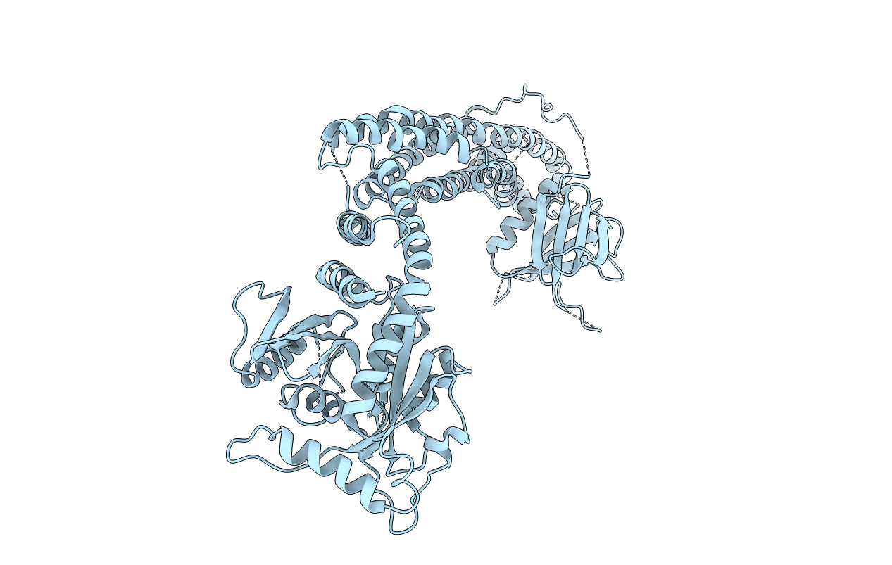
Crystal structure of nucleotide-free human dynamin1
Biological Source:
Source Organism:
Homo sapiens (Taxon ID: 9606)
Host Organism:
Method Details:
Experimental Method:
Resolution:
3.70 Å
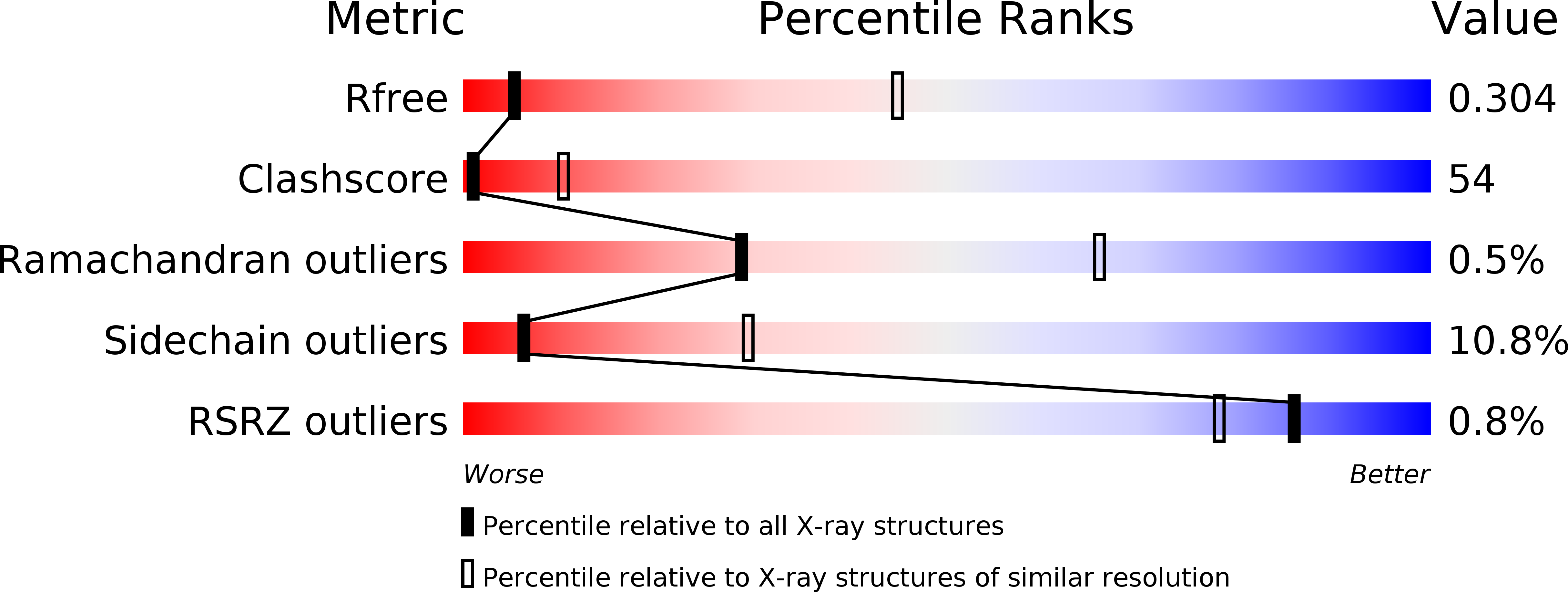
R-Value Free:
0.33
R-Value Work:
0.28
R-Value Observed:
0.29
Space Group:
C 2 2 21
Macromolecular Entities
Structures with similar UniProt ID
Protein Blast
Polymer Type:polypeptide(L)
Molecule:Dynamin-1
Gene (Uniprot):DNM1
Mutagens:I395A,H396A,G397A,I398A,R399A,K562E
Chain IDs:A
Chain Length:743
Number of Molecules:1
Biological Source:Homo sapiens
Ligand Molecules
Primary Citation
Crystal structure of nucleotide-free dynamin.
Nature 477 556 560 (2011)
PMID: 21927000 DOI: 10.1038/nature10369

Abstact

Dynamin is a mechanochemical GTPase that oligomerizes around the neck of clathrin-coated pits and catalyses vesicle scission in a GTP-hydrolysis-dependent manner. The molecular details of oligomerization and the mechanism of the mechanochemical coupling are currently unknown. Here we present the crystal structure of human dynamin 1 in the nucleotide-free state with a four-domain architecture comprising the GTPase domain, the bundle signalling element, the stalk and the pleckstrin homology domain. Dynamin 1 oligomerized in the crystals via the stalks, which assemble in a criss-cross fashion. The stalks further interact via conserved surfaces with the pleckstrin homology domain and the bundle signalling element of the neighbouring dynamin molecule. This intricate domain interaction rationalizes a number of disease-related mutations in dynamin 2 and suggests a structural model for the mechanochemical coupling that reconciles previous models of dynamin function.

Legend

Protein

Chemical

Disease

Primary Citation of related structures