3SN6 image
Deposition Date 2011-06-28
Release Date 2011-07-20
Last Version Date 2024-11-20
Entry Detail
PDB ID:
3SN6
Title:
Crystal structure of the beta2 adrenergic receptor-Gs protein complex
Biological Source:
Source Organism:
Bos taurus (Taxon ID: 9913)
Rattus norvegicus (Taxon ID: 10116)
Enterobacteria phage T4 (Taxon ID: 10665)
Homo sapiens (Taxon ID: 9606)
Lama glama (Taxon ID: 9844)
Host Organism:
Method Details:
Experimental Method:
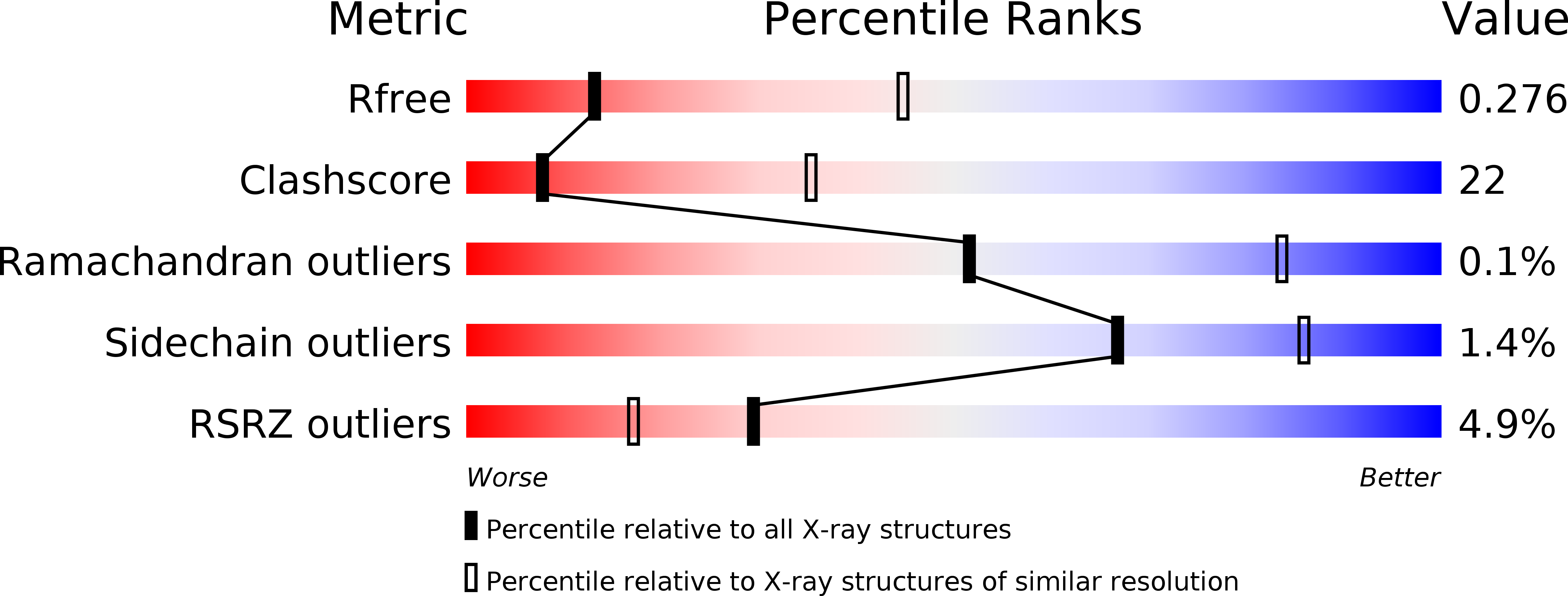
Resolution:
3.20 Å
R-Value Free:
0.27
R-Value Work:
0.22
R-Value Observed:
0.22
Space Group:
P 1 21 1
Macromolecular Entities
Structures with similar UniProt ID
Protein Blast
Polymer Type:polypeptide(L)
Molecule:Guanine nucleotide-binding protein G(s) subunit alpha isoforms short
Gene (Uniprot):GNAS
Mutagens:G72S
Chain IDs:A
Chain Length:380
Number of Molecules:1
Biological Source:Bos taurus
Structures with similar UniProt ID
Protein Blast
Polymer Type:polypeptide(L)
Molecule:Guanine nucleotide-binding protein G(I)/G(S)/G(T) subunit beta-1
Gene (Uniprot):Gnb1
Mutagens:M1Q
Chain IDs:B
Chain Length:351
Number of Molecules:1
Biological Source:Rattus norvegicus
Structures with similar UniProt ID
Protein Blast
Polymer Type:polypeptide(L)
Molecule:Guanine nucleotide-binding protein G(I)/G(S)/G(O) subunit gamma-2
Gene (Uniprot):GNG2
Chain IDs:C (auth: G)
Chain Length:68
Number of Molecules:1
Biological Source:Bos taurus
Polymer Type:polypeptide(L)
Molecule:Camelid antibody VHH fragment
Chain IDs:E (auth: N)
Chain Length:138
Number of Molecules:1
Biological Source:Lama glama
Protein Blast
Polymer Type:polypeptide(L)
Molecule:Endolysin,Beta-2 adrenergic receptor
Gene (Uniprot):E, ADRB2
Mutagens:C54T,C97A,M96T,M98T,N187E
Chain IDs:D (auth: R)
Chain Length:514
Number of Molecules:1
Biological Source:Enterobacteria phage T4, Homo sapiens
Ligand Molecules
Primary Citation

Abstact

G protein-coupled receptors (GPCRs) are responsible for the majority of cellular responses to hormones and neurotransmitters as well as the senses of sight, olfaction and taste. The paradigm of GPCR signalling is the activation of a heterotrimeric GTP binding protein (G protein) by an agonist-occupied receptor. The β(2) adrenergic receptor (β(2)AR) activation of Gs, the stimulatory G protein for adenylyl cyclase, has long been a model system for GPCR signalling. Here we present the crystal structure of the active state ternary complex composed of agonist-occupied monomeric β(2)AR and nucleotide-free Gs heterotrimer. The principal interactions between the β(2)AR and Gs involve the amino- and carboxy-terminal α-helices of Gs, with conformational changes propagating to the nucleotide-binding pocket. The largest conformational changes in the β(2)AR include a 14 Å outward movement at the cytoplasmic end of transmembrane segment 6 (TM6) and an α-helical extension of the cytoplasmic end of TM5. The most surprising observation is a major displacement of the α-helical domain of Gαs relative to the Ras-like GTPase domain. This crystal structure represents the first high-resolution view of transmembrane signalling by a GPCR.

Legend

Protein

Chemical

Disease

Primary Citation of related structures
Feedback Form
Name
Email
Institute
Feedback