3SJH image
Deposition Date 2011-06-21
Release Date 2012-01-25
Last Version Date 2023-09-13
Entry Detail
PDB ID:
3SJH
Title:
Crystal Structure of a chimera containing the N-terminal domain (residues 8-29) of drosophila Ciboulot and the C-terminal domain (residues 18-44) of bovine Thymosin-beta4, bound to G-actin-ATP-Latrunculin A
Biological Source:
Source Organism:
Host Organism:
Method Details:
Experimental Method:
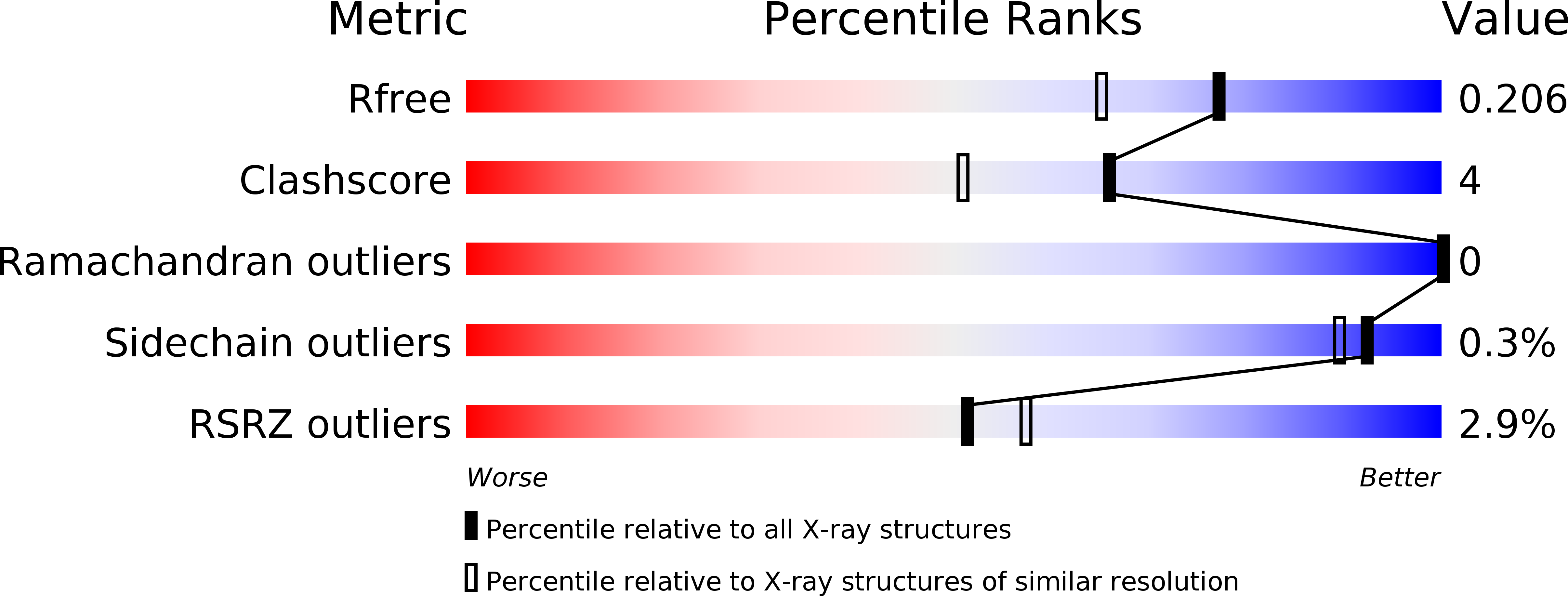
Resolution:
1.75 Å
R-Value Free:
0.19
R-Value Work:
0.16
R-Value Observed:
0.17
Space Group:
P 21 21 21
Macromolecular Entities
Polymer Type:polypeptide(L)
Molecule:Actin, alpha skeletal muscle
Gene (Uniprot):ACTA1
Chain IDs:A
Chain Length:375
Number of Molecules:1
Biological Source:Oryctolagus cuniculus
Polymer Type:polypeptide(L)
Molecule:Ciboulot/Thymosin beta-4 chimeric protein
Gene (Uniprot):cib, TMSB4
Chain IDs:B
Chain Length:54
Number of Molecules:1
Biological Source:Drosophila melanogaster, Bos taurus
Primary Citation
How a single residue in individual beta-thymosin/WH2 domains controls their functions in actin assembly
Embo J. 31 1000 1013 (2012)
PMID: 22193718 DOI: 10.1038/emboj.2011.461

Abstact

β-Thymosin (βT) and WH2 domains are widespread, intrinsically disordered actin-binding peptides that display significant sequence variability and different regulations of actin self-assembly in motile and morphogenetic processes. Here, we reveal the structural mechanisms by which, in their 1:1 stoichiometric complexes with actin, they either inhibit assembly by sequestering actin monomers like Thymosin-β4, or enhance motility by directing polarized filament assembly like Ciboulot βT. We combined mutational, functional or structural analysis by X-ray crystallography, SAXS (small angle X-ray scattering) and NMR on Thymosin-β4, Ciboulot, TetraThymosinβ and the long WH2 domain of WASP-interacting protein. The latter sequesters G-actin with the same molecular mechanisms as Thymosin-β4. Functionally different βT/WH2 domains differ by distinct dynamics of their C-terminal half interactions with G-actin pointed face. These C-terminal interaction dynamics are controlled by the strength of electrostatic interactions with G-actin. At physiological ionic strength, a single salt bridge with actin located next to their central LKKT/V motif induces G-actin sequestration in both isolated long βT and WH2 domains. The results open perspectives for elucidating the functions of βT/WH2 domains in other modular proteins.

Legend

Protein

Chemical

Disease

Primary Citation of related structures