3SIX image
Deposition Date 2011-06-20
Release Date 2012-02-08
Last Version Date 2023-09-13
Entry Detail
PDB ID:
3SIX
Keywords:
Title:
Crystal structure of NodZ alpha-1,6-fucosyltransferase soaked with GDP-fucose
Biological Source:
Source Organism:
Host Organism:
Method Details:
Experimental Method:
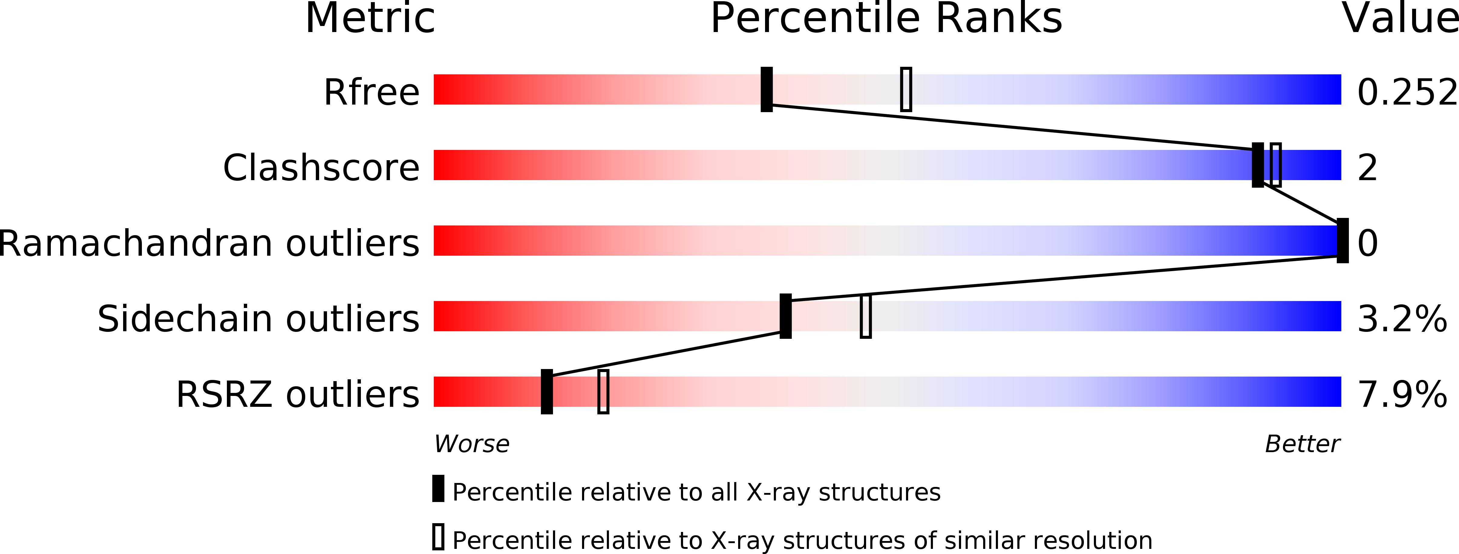
Resolution:
2.35 Å
R-Value Free:
0.25
R-Value Work:
0.21
R-Value Observed:
0.21
Space Group:
P 65 2 2
Macromolecular Entities
Structures with similar UniProt ID
Protein Blast
Polymer Type:polypeptide(L)
Molecule:Nodulation fucosyltransferase NodZ
Gene (Uniprot):nodZ
Chain IDs:A
Chain Length:330
Number of Molecules:1
Biological Source:Bradyrhizobium sp.
Primary Citation

Abstact

Rhizobial NodZ α1,6-fucosyltransferase (α1,6-FucT) catalyzes the transfer of the fucose (Fuc) moiety from guanosine 5'-diphosphate-β-L-fucose to the reducing end of the chitin oligosaccharide core during Nod-factor (NF) biosynthesis. NF is a key signalling molecule required for successful symbiosis with a legume host for atmospheric nitrogen fixation. To date, only two α1,6-FucT structures have been determined, both without any donor or acceptor molecule that could highlight the structural background of the catalytic mechanism. Here, the first crystal structures of α1,6-FucT in complex with its substrate GDP-Fuc and with GDP, which is a byproduct of the enzymatic reaction, are presented. The crystal of the complex with GDP-Fuc was obtained through soaking of native NodZ crystals with the ligand and its structure has been determined at 2.35 Å resolution. The fucose residue is exposed to solvent and is disordered. The enzyme-product complex crystal was obtained by cocrystallization with GDP and an acceptor molecule, penta-N-acetyl-L-glucosamine (penta-NAG). The structure has been determined at 1.98 Å resolution, showing that only the GDP molecule is present in the complex. In both structures the ligands are located in a cleft formed between the two domains of NodZ and extend towards the C-terminal domain, but their conformations differ significantly. The structures revealed that residues in three regions of the C-terminal domain, which are conserved among α1,2-, α1,6- and protein O-fucosyltransferases, are involved in interactions with the sugar-donor molecule. There is also an interaction with the side chain of Tyr45 in the N-terminal domain, which is very unusual for a GT-B-type glycosyltransferase. Only minor conformational changes of the protein backbone are observed upon ligand binding. The only exception is a movement of the loop located between strand βC2 and helix αC3. In addition, there is a shift of the αC3 helix itself upon GDP-Fuc binding.

Legend

Protein

Chemical

Disease

Primary Citation of related structures