3SID image
Deposition Date 2011-06-17
Release Date 2011-08-31
Last Version Date 2024-10-30
Entry Detail
PDB ID:
3SID
Keywords:
Title:
Crystal structure of oxidized Symerythrin from Cyanophora paradoxa, azide adduct at 50% occupancy
Biological Source:
Source Organism:
Host Organism:
Method Details:
Experimental Method:
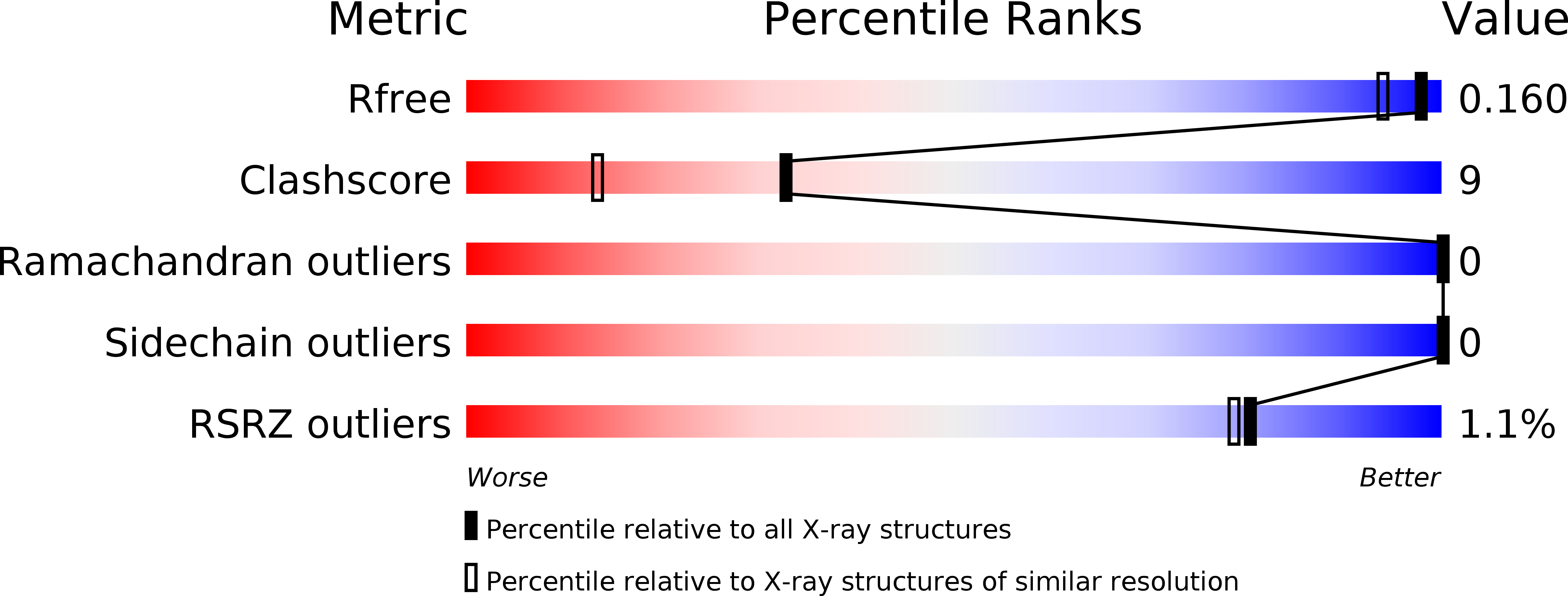
Resolution:
1.40 Å
R-Value Free:
0.15
R-Value Work:
0.11
R-Value Observed:
0.11
Space Group:
P 32
Macromolecular Entities
Polymer Type:polypeptide(L)
Molecule:Symerythrin
Chain IDs:A, B
Chain Length:180
Number of Molecules:2
Biological Source:Cyanophora paradoxa
Primary Citation
Symerythrin structures at atomic resolution and the origins of rubrerythrins and the ferritin-like superfamily.
J.Mol.Biol. 413 177 194 (2011)
PMID: 21872605 DOI: 10.1016/j.jmb.2011.08.019

Abstact

Rubrerythrins are diiron-containing peroxidases that belong to the ferritin-like superfamily (FLSF). Here, we describe the structures of symerythrin, a novel rubrerythrin variant from the oxygenic phototroph Cyanophora paradoxa, at 1.20-1.40 Å resolution in three different states: diferric, azide-bound diferric and chemically reduced. The symerythrin metallocenter has a unique eighth ligating residue compared to rubrerythrin-an additional glutamate inserted into helix A of the four-helix bundle that resides on a π-helical segment. Otherwise, the diferric metallocenter structure is highly similar to that of characterized rubrerythrins. Azide binds the diferric center in a μ-1,1 orientation similar to how peroxide binds to diferric rubrerythrin. The structure of the diferrous metallocenter shows heterogeneity that we ascribe to the acidic pH of the crystals. In what we consider the neutral pH conformation, reduction causes a 2.0-Å shift in Fe1 and the toggling of a Glu to a His ligand, as seen with rubrerythrins. The function of symerythrin remains unknown, but preliminary tests showing oxidase and peroxidase activities and the similarities of its metallocenter to other rubrerythrins suggest similar functionalities between the two despite the additional ligating glutamate in symerythrin. Of particular interest is the high internal symmetry of symerythrin, which supports the notion that its core four-helix bundle was formed by the gene duplication and fusion of a two-helix peptide. Sequence comparisons with another family in the FLSF that also has notable internal symmetry provide compelling evidence that, contrary to previous assumptions, there have been multiple gene fusion events that have generated the single-chain FLSF fold.

Legend

Protein

Chemical

Disease

Primary Citation of related structures