3SI9 image
Deposition Date 2011-06-17
Release Date 2011-06-29
Last Version Date 2023-09-13
Entry Detail
PDB ID:
3SI9
Keywords:
Title:
Crystal structure of Dihydrodipicolinate Synthase from Bartonella Henselae
Biological Source:
Source Organism:
Host Organism:
Method Details:
Experimental Method:
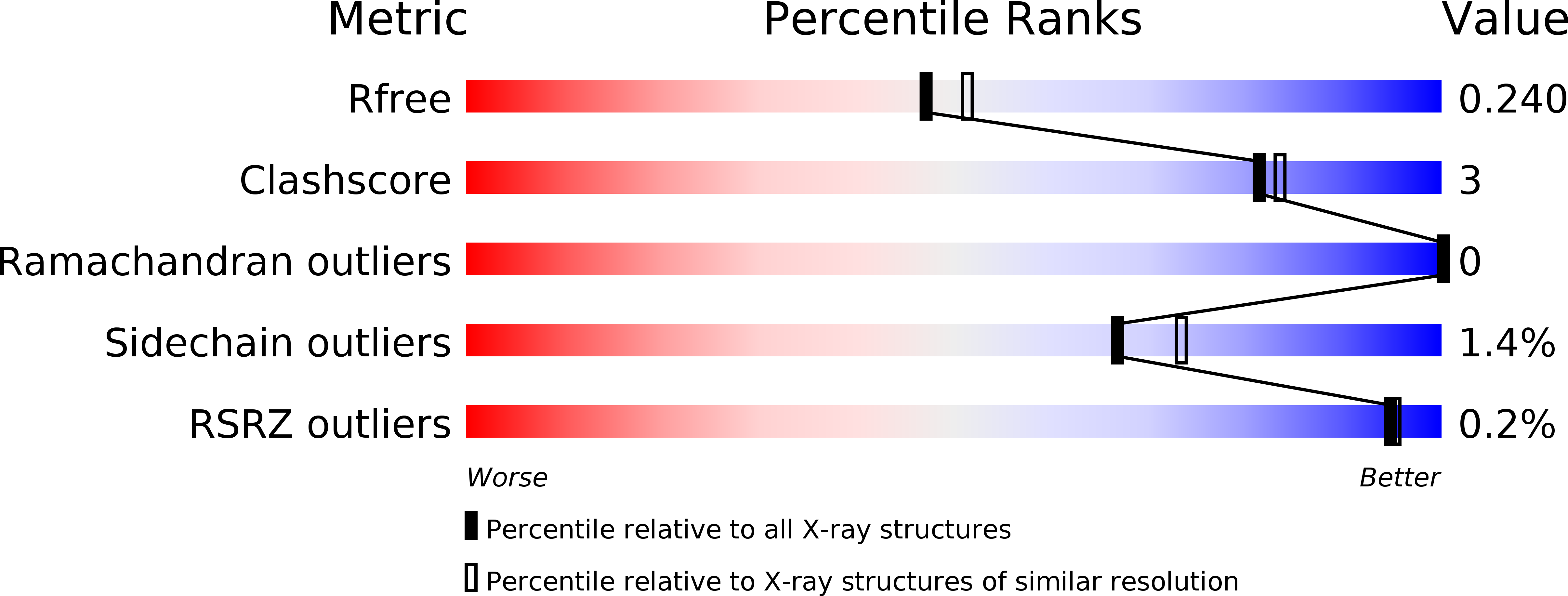
Resolution:
2.10 Å
R-Value Free:
0.23
R-Value Work:
0.18
R-Value Observed:
0.18
Space Group:
P 21 21 21
Macromolecular Entities
Polymer Type:polypeptide(L)
Molecule:Dihydrodipicolinate synthase
Gene (Uniprot):dapA
Chain IDs:A, B, C, D
Chain Length:315
Number of Molecules:4
Biological Source:Bartonella henselae
Ligand Molecules
Primary Citation
Cloning, expression, purification, crystallization and X-ray diffraction analysis of dihydrodipicolinate synthase from the human pathogenic bacterium Bartonella henselae strain Houston-1 at 2.1 angstrom resolution.
Acta Crystallogr F Struct Biol Commun 72 2 9 (2016)
PMID: 26750477 DOI: 10.1107/S2053230X15023213

Abstact

The enzyme dihydrodipicolinate synthase catalyzes the committed step in the synthesis of diaminopimelate and lysine to facilitate peptidoglycan and protein synthesis. Dihydrodipicolinate synthase catalyzes the condensation of L-aspartate 4-semialdehyde and pyruvate to synthesize L-2,3-dihydrodipicolinate. Here, the cloning, expression, purification, crystallization and X-ray diffraction analysis of dihydrodipicolinate synthase from the pathogenic bacterium Bartonella henselae, the causative bacterium of cat-scratch disease, are presented. Protein crystals were grown in conditions consisting of 20%(w/v) PEG 4000, 100 mM sodium citrate tribasic pH 5.5 and were shown to diffract to ∼2.10 Å resolution. They belonged to space group P212121, with unit-cell parameters a = 79.96, b = 106.33, c = 136.25 Å. The final R values were Rr.i.m. = 0.098, Rwork = 0.183, Rfree = 0.233.

Legend

Protein

Chemical

Disease

Primary Citation of related structures