3SHI image
Deposition Date 2011-06-16
Release Date 2011-09-21
Last Version Date 2023-09-13
Entry Detail
PDB ID:
3SHI
Keywords:
Title:
Crystal structure of human MMP1 catalytic domain at 2.2 A resolution
Biological Source:
Source Organism:
Homo sapiens (Taxon ID: 9606)
Host Organism:
Method Details:
Experimental Method:
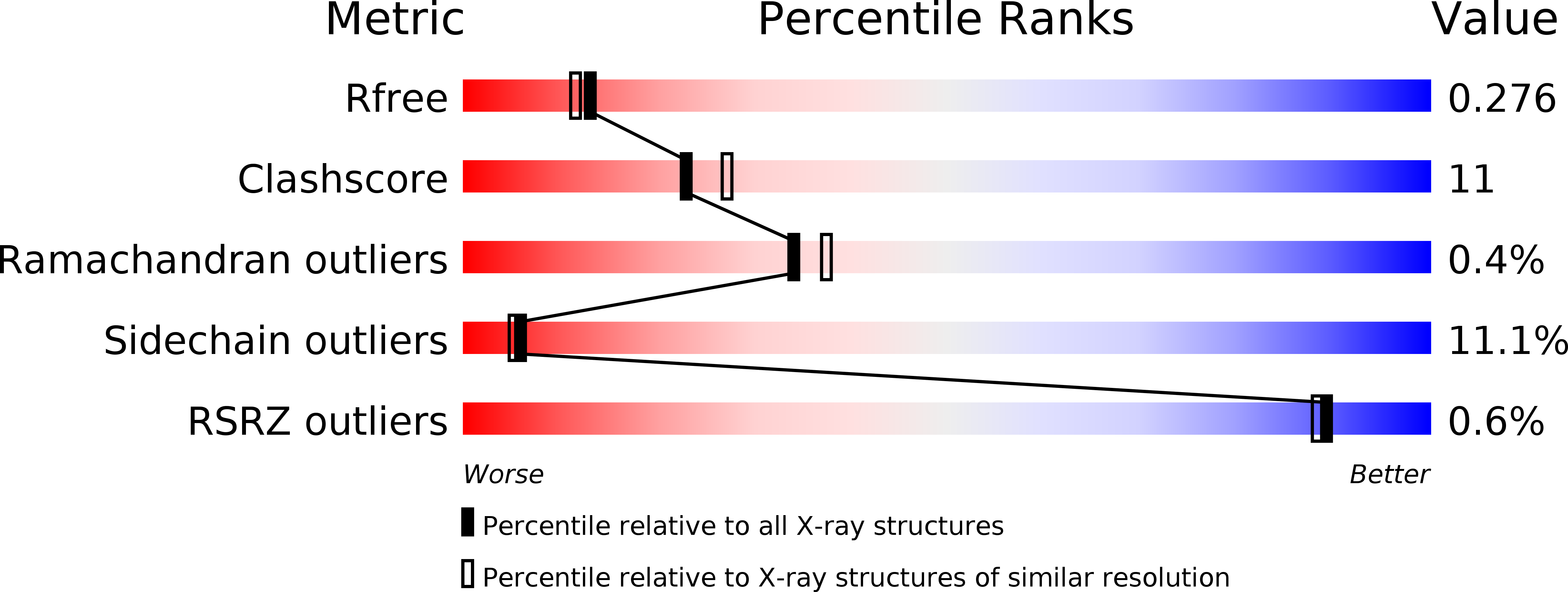
Resolution:
2.20 Å
R-Value Free:
0.27
R-Value Work:
0.20
R-Value Observed:
0.21
Space Group:
C 1 2 1
Macromolecular Entities
Polymer Type:polypeptide(L)
Molecule:Interstitial collagenase
Gene (Uniprot):MMP1
Chain IDs:A, B (auth: G), C (auth: M)
Chain Length:156
Number of Molecules:3
Biological Source:Homo sapiens
Primary Citation

Abstact

Pseudocontact shifts (pcs) and paramagnetic residual dipolar couplings (rdc) provide structural information that can be used to assess the adequacy of a crystallographic structure to represent the solution structure of a protein. This can be done by attaching a lanthanide binding tag to the protein. There are cases in which only local rearrangements are sufficient to match the NMR data and cases where significant secondary structure or domain rearrangements from the solid state to the solution state are needed. We show that the two cases are easily distinguishable. Whereas the use of solution restraints in the latter case is described in the literature, here we deal with how to obtain a better model of the solution structure in a case (the catalytic domain of the matrix metalloproteinase MMP-1) of the former class.

Legend

Protein

Chemical

Disease

Primary Citation of related structures