Deposition Date
2011-06-16
Release Date
2011-08-24
Last Version Date
2024-11-06
Entry Detail
PDB ID:
3SHF
Keywords:
Title:
Crystal structure of the R265S mutant of full-length murine Apaf-1
Biological Source:
Source Organism(s):
Mus musculus (Taxon ID: 10090)
Expression System(s):
Method Details:
Experimental Method:
Resolution:
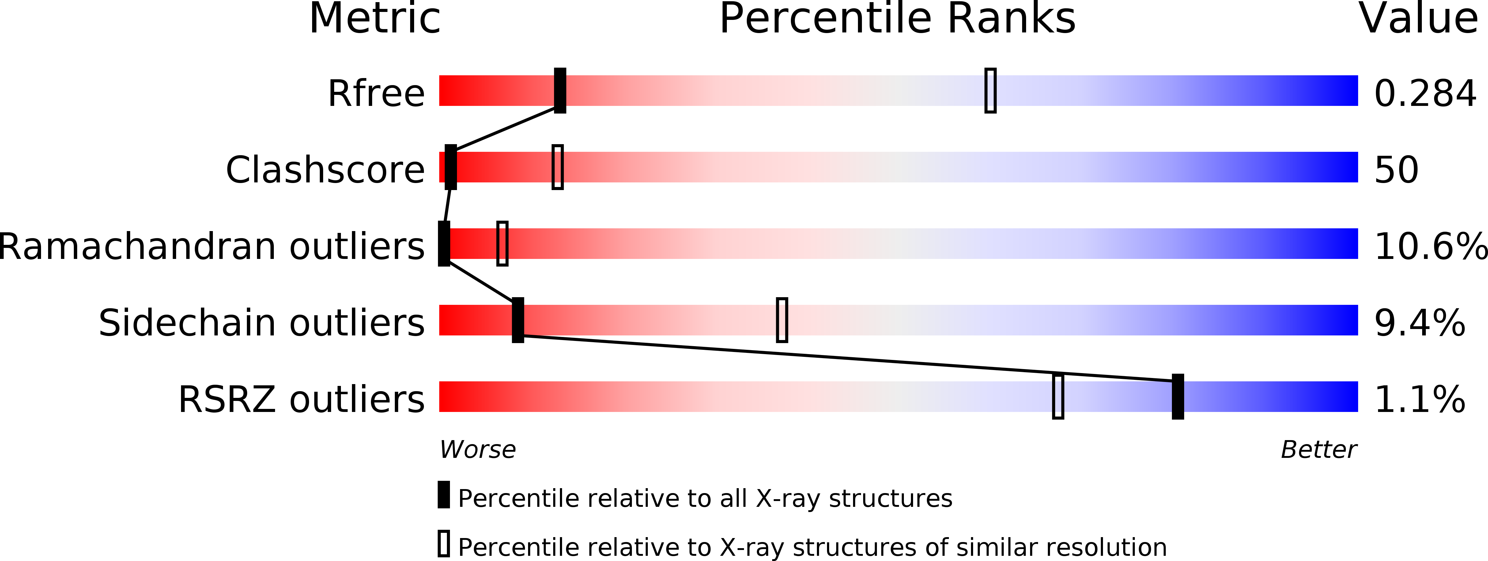
3.55 Å
R-Value Free:
0.30
R-Value Work:
0.22
Space Group:
P 21 21 21